Epitheloidsarkom der Cervix uteri

Epithelioid
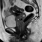
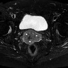
sarcoma of the uterine cervix. Sagittal (a), axial (b) T2-weighted images showed moderate-to-high signal intensity mass (*) at lower cervix (3 cm craniocaudal diameter), which obliterated the vaginal fornices. Note spared upper cervix, normal-sized uterine body and fundus.

Epithelioid
sarcoma of the uterine cervix. Axial (b & fat-suppressed c) T2-weighted images confirmed predominantly exophytic lower cervical mass (*) with moderate-to-high signal intensity, 52x36mm axial diameters, thinned disrupted outer stroma consistent with parametrial infiltration.

Epithelioid
sarcoma of the uterine cervix. Axial (b & fat-suppressed c) T2-weighted images confirmed predominantly exophytic lower cervical mass (*) with moderate-to-high signal intensity, 52x36mm axial diameters. Note absent pelvic lymphadenopathies and effusion in peritoneal cul-de-sac.

Epithelioid
sarcoma of the uterine cervix. On precontrast T1-weighted images the lower cervical exophytic mass (*) showed solid-type intermediate signal intensity. Note absent pelvic lymphadenopathies and effusion in peritoneal cul-de-sac.

Epithelioid
sarcoma of the uterine cervix. High (800) b-value diffusion-weighted images (e) showed visually hyperintense signal in the lobulated, well demarcated lower cervical mass (*), reflecting restricted diffusion, with low signal on corresponding apparent diffusion coefficient (ADC) map (f).

Epithelioid
sarcoma of the uterine cervix. In the apparent diffusion coefficient (ADC) map the lobulated lower cervical mass (*) showed low signal from restricted diffusion, with measured ADC values of approximately 0.70-0.75x10⁻³ mm2/s in its peripheral portions.

Epithelioid
sarcoma of the uterine cervix. Limited to axial T2-weighted images because of claustrophobia, repeated MRI showed stable size and thickness of lower cervical mass (*) compared to Fig. 1, without lymphadenopathies and peritoneal effusion.
Epitheloidsarkom der Cervix uteri
Das Epitheloidsarkom ist ein seltenes, aber aggressives Weichteilsarkom mit epitheloider Zellmorphologie und häufig ungünstiger Prognose aufgrund von Lokalrezidiven und Metastasen. Häufige Lokalisationen sind die distalen Extremitäten, jedoch sind auch andere Fälle zum Beispiel an den Organen des Beckens beschrieben.
Siehe auch:

 Assoziationen und Differentialdiagnosen zu Epitheloidsarkom der Cervix uteri:


