esophageal foreign body

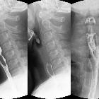
Fishbone
pierced in the upper esophagus.Left image during swallowing contrast medium, right image after swallow only dimly visible.
Steckengebliebenes
Stück einer Karotte im oberen Ösophagus knapp unterhalb des oberen Sphinkters.
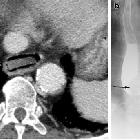
Imaging of
the oesophagus: beyond cancer. Foreign body impaction. a Contrast-enhanced CT of the chest in soft tissue window shows a square-shaped opacity (piece of carrot) impacted within the thoracic oesophagus just above the level of the diaphragm. b Esophagram in AP projection in another patient with dysphagia demonstrates a round filling defect within the midthoracic oesophagus. This was endoscopically confirmed to be an impacted hot dog


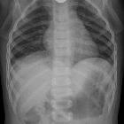
Verschluckter
und im oberen Ösophagus steckengebliebener Hühnerknochen in der Breischluckuntersuchung als schräg liegende Kontrastmittelaussparung zu erkennen.

Imaging of
the oesophagus: beyond cancer. Complication of food impaction with tear of oesophagus during chicken bone extraction. a Pre-contrast axial image of the thorax demonstrates a markedly enlarged oesophagus which is high in density (*) when compared to the adjacent blood pool in the aorta (findings consistent with hematoma). b Post-contrast imaging of the same patient demonstrates areas of active contrast extravasation (arrows) within a markedly enlarged and abnormal appearing oesophagus. Clinical note, patient subsequently had multiple episodes of hematemesis

Gastrointestinal
perforation: clinical and MDCT clues for identification of aetiology. 77-year-old patient with prolonged dysphagia. Sagittal reformatted image in bone window demonstrates a swallowed perforating denture (*) impinging on the upper oesophagus. There is superficial (arrows) and deep (arrowheads) cervical emphysema
ösophagealer Fremdkörper
esophageal foreign body
Siehe auch:
- Fremdkörper
- Hühnerknochen verschluckt
- verschluckte Fremdkörper
- Fischgräte verschluckt
- Fremdkörperimpaktation
und weiter:


 Assoziationen und Differentialdiagnosen zu ösophagealer Fremdkörper:
