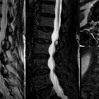
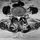
Facettengelenkszyste LWS

Facettengelenksarthrose
mit Facettengelenks-Zyste von rechts nach intraspinal mit hochgradiger spinaler Enge.

Intraspinale,
epidurale, eingeblutete Synovialzyste Facettengelenk mit akuter Klinik mit Schmerz und Kaudasymptomatik. Oben sagittal T1 nativ (hell !), T2, STIR, unten T2 axial , T1 KM FS axial und sagittal.

Synovialiszyste
des Facettengelenks rechts in der Etage LWK 1/2 mit nur geringer Pelottierung des Duralsackes.

Facettengelenksarthrose
mit Facettengelenks-Zyste nach dorsal. Es findet sich vermehrt Flüssigkeit im Gelenkspalt, links mehr als rechts. Die Zyste schließt sich dorsal links an.

Spectrum of
MRI features of ganglion and synovial cysts. a-c. Lumbar facet synovial cyst in an 82-year-old woman presenting with subacute left lumbar radiculopathy and neurogenic claudication. Sagittal T2-weighted MRI (a) shows a slightly hyperintense cystic lesion posteriorly to the L3/L4 disc (arrow), as well as grade 1 degenerative spondylolisthesis at L4/L5. The axial view (b) clearly demonstrates the extradural location of the lesion (dashed arrow) arising from the left L3/L4 degenerated facet joint, which presents synovial effusion (asterisk). Note in both axial and coronal (c) views the displacement of the thecal sac and the left L4 nerve root (arrows) toward the right, due to compression by the cyst (dashed arrows). The partial T2-hypointensity, more evident in image c, might correspond to high-protein content or previous internal bleeding

Spectrum of
MRI features of ganglion and synovial cysts. a, b. Lumbar facet synovial cyst in a 50-year-old man with a history of spinal surgery due to spondylolisthesis 20 years earlier, presenting with low back pain. Axial (a) and sagittal (b) T2-weighted images show a mildly hyperintense extradural rounded lesion (dashed arrows) arising from the right L4/L5 facet joint, which presents marked degenerative changes and fluid (asterisk). Note the compression of the thecal sac, displaced posteriorly (arrow in b) and to the left side (arrow in a). Case courtesy of Dr. Carlos Casimiro. Neuroradiology Department, Centro Hospitalar de Lisboa Norte. Lisbon, Portugal
Facettengelenkszyste LWS
Siehe auch:
- Wurzeltaschenzysten
- Synovialzyste
- Spondylarthrose
- intraspinale zystische Läsionen
- Synovialzysten der Facettengelenke
und weiter:

 Assoziationen und Differentialdiagnosen zu Facettengelenkszyste LWS:
