fatty metamorphosis of the liver

Multifocal
nodular hepatic steatosis. CT showed more nodular hepatic lesions than those visualized in previous CTs. The radiologic appearance of these lesions had not changed but their size had significantly increased.

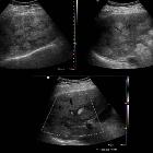
Multifocal
nodular hepatic steatosis. Multiple hyperechoic and well-defined nodular lesions which corresponded to focal nodular steatosis. Core-needle biopsy confirmed the presence of macrovesicular steatosis within these lesions.

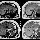
Multifocal
nodular hepatic steatosis. Multiple nodular hepatic lesions clearly depicted on T1-weighted out-of-phase images. These lesions were not visible on T1 weighted in-phase images.
Multifocal
nodular hepatic steatosis. Multifocal nodular hepatic lesions, mainly hypodense (red arrows) associated to some diffuse hypodense hepatic areas (black arrows). Note the splenic haematoma and an artefact in the lower pole of the spleen (embolization).J.A Prat-Matifoll, Vall Hebron Hospital, Radiology Department
Multifocal
nodular hepatic steatosis. Multiple nodular lesions were clearly depicted on a T1-weighted out-of-phase image but were slightly hyperintense on a T2-weighted image. These lesions were not visible in diffusion or ADC. No contrast enhancement was observed.J.A Prat-Matifoll, Vall Hebron Hospital, Radiology Department


 Assoziationen und Differentialdiagnosen zu fatty metamorphosis of the liver:


