Fischgräte

Fishbone
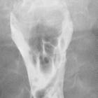
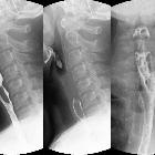
pierced in the upper esophagus.Left image during swallowing contrast medium, right image after swallow only dimly visible.

Ingested
bones • Ingested fish bone with sealed duodenal perforation - Ganzer Fall bei Radiopaedia

Gastrointestinal
perforation: clinical and MDCT clues for identification of aetiology. 70-year-old patient presenting with fever and right upper quadrant pain for 3 days. Axial contrast-enhanced image demonstrates a gas-containing liver abscess (open arrow) and a hyperdense foreign body in the hepatogastric ligament representing a penetrating fishbone (arrowhead)

Is that... a
Meckel"s diverticulum?!. Coronal (a) and axial (b) non-enhanced CT images showing linear hyperdense image measuring about 5cm, apparently located in the terminal ileum loop and perforating the wall. Focal inflammatory changes were also present.

Is that... a
Meckel"s diverticulum?!. Linear highly hyperchogenic image within an apparent terminal ileum loop, in a fixed transverse position, compatible with a foreign body.

Is that... a
Meckel"s diverticulum?!. Coronal (a) and axial (b) non-enhanced CT images showing linear hyperdense image measuring about 5cm, apparently located in the terminal ileum loop and perforating the wall. Focal inflammatory changes were also present.

Unconscious
ingestion of fishbone detected on MDCT.. Axial unenhanced CT image showing the hyperdense structure, excluding the presence of contrast extravasation.

Unconscious
ingestion of fishbone detected on MDCT.. Coronal reformatted image showing the thickened loop, the hyperdense structure (arrow) along with the mesenteric fat stranding.

Unconscious
ingestion of fishbone detected on MDCT.. Sagittal reformatted image showing the hyperdense structure (arrow).

Unconscious
ingestion of fishbone detected on MDCT.. Axial contrast-enhanced CT image showing the fat stranding associated with the thickened jejunal loop.

Unconscious
ingestion of fishbone detected on MDCT.. Axial contrast-enhanced CT image showing a focally thickened jejunal loop with marked fat stranding of the adjacent mesenteric fat. Note a hyperdense linear structure situated at the loop’s wall.

Ingested
bones • Neck foreign body - fish bone - Ganzer Fall bei Radiopaedia

The
anatomical compartments and their connections as demonstrated by ectopic air. Local sources of pneumomediastinum. a, b Spontaneous pneumomediastinum due to prolonged and forceful Valsalva manoeuvres in 21-year-old man. Axial CT scan with “lung window” demonstrates the Macklin effect: air is seen along the perivascular and peribronchial fascial sheaths (a), continuous to the mediastinum (red star) (b). c Oesophageal perforation by fishbone complicated by mediastinitis in a 61-year-old woman. Axial CT scan after oral iodinated contrast ingestion, distending the oesophagus (red square); extraluminal air bubbles (red arrows) are seen in mediastinum together with increased mediastinal fat attenuation, as well as multiple reactive lymph nodes. Normal permeable airways are seen (blue triangle) at level of carina

Ingested
bones • Ingested foreign body - fish bone - Ganzer Fall bei Radiopaedia

Pneumomediastinum
• Pneumomediastinum from ingested fish bone - Ganzer Fall bei Radiopaedia

Ingested
bones • Fish bone impaction into the pharyngeal wall - Ganzer Fall bei Radiopaedia

Ingested
bones • Distal esophageal perforation by a fishbone - Ganzer Fall bei Radiopaedia

Ingested
bones • Liver punctured by fishbone - Ganzer Fall bei Radiopaedia

Ingested
bones • Foreign body - fishbone - Ganzer Fall bei Radiopaedia

Ingested
bones • Small bowel perforation - fish bone - Ganzer Fall bei Radiopaedia

Ingested
bones • Fish bone stuck in vallecula - Ganzer Fall bei Radiopaedia

Laparoscopic
removal of an ingested fish bone that penetrated the stomach and was embedded in the pancreas: a case report. Coronal view of a computed tomography (CT) image showing a linear, hyperdense, foreign body (arrow), which appeared to penetrate through the posterior wall of the gastric antrum (a). CT image showing the foreign body (arrow) and the pancreas (arrowhead) (b)

Duodenal
perforation secondary to an ingested fish bone. CT scan showed a lineal high-density structure measuring 23 mm located at the duodenal wall and protruding towards the omental fat. A small bubble of air and surrounding inflammatory changes were also depicted.

Gastrointestinal
perforation: clinical and MDCT clues for identification of aetiology. 78-year-old patient. Contrast-enhanced axial image demonstrates a linear hyperdense foreign body partly protruding through the ileal wall, representing a fishbone (arrow). Note the concentric wall thickening of the affected loop, few adjacent gas bubbles (arrowhead) and pneumoperitoneum (p)

 Assoziationen und Differentialdiagnosen zu Fischgräte verschluckt:

