hämorrhagischer Infarkt in Makroadenom der Hypophyse

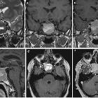
Pituitary
apoplexy: an update on clinical and imaging features. MRI performed in a patient with acute headache, mydriasis and visual impairment shows acute/early subacute phase haemorrhagic infarction within a pituitary macroadenoma. The intrasellar mass appears slightly hyperintense on unenhanced T1WI (axial, a; coronal, b; sagittal, c) with areas of hypointense signal on T2WI (axial, d; coronal, e) because of the presence of deoxyhaemoglobin. Coronal T1 contrast-enanched images (f) show left cavernous sinus involvement, probably leading to visual symptoms
Pituitary
apoplexy: an update on clinical and imaging features. Large pituitary macroadenoma with MRI signs of subacute haemorrhage. In this phase, haemorrhage appears hyperintense on both T1WI (b) and T2WI (a). In the T2WI (a, e), thickening of the sphenoid mucosa (arrow) is evident as well. This sign is highly specific for pituitary apoplexy. On contrast-enhanced MRI (c, d, f) the residual pituitary gland enhances (arrow in c compared to b). Note the optic pathway compression, more evident on T2WI
hämorrhagischer Infarkt in Makroadenom der Hypophyse
Siehe auch:


 Assoziationen und Differentialdiagnosen zu hämorrhagischer Infarkt in Makroadenom der Hypophyse: