Hematuria (adult)
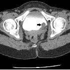
Hematuria occurs when blood enters the urinary collecting system and is excreted in the urine. There are many etiologies for hematuria, and they range from benign and transient to gravely concerning. Hematuria can derive from the kidneys, ureters, bladder, prostate (in men), or urethra. Imaging can often be useful to determine the source.
Pathology
Hematuria can be considered in two main forms:
- frank (macroscopic) hematuria
- microscopic hematuria
Frank hematuria
Frank hematuria is considered >1 mL of blood in the urine. Red-colored urine (more red than brown) or clots in the urine is suggestive of a lower urinary tract source.
Microscopic hematuria
Microscopic hematuria may be either symptomatic or asymptomatic and is defined as ≥3 red blood cells per high-powered field (HPF) on 2 out of 3 urinalysis specimens .
Treatment and prognosis
Frank hematuria is almost always evaluated clinically, with cystoscopy, and with imaging (e.g. CT urogram).
Imaging workup for microscopic hematuria is controversial. Some academic societies advocate that in the absence of a known benign etiology (such as vigorous exercise, infection, or menstruation), then a CT urogram is warranted . If the patient has known renal disease then an ultrasound of the kidneys and bladder may be more appropriate.
Some feel that the imaging workup in the setting of microscopic hematuria has a high negative rate for malignancy, and suggest that instead of imaging all patients with microscopic hematuria, imaging should be reserved for higher-risk subsets .
Microscopic hematuria in children has a different treatment algorithm.
Siehe auch:


 Assoziationen und Differentialdiagnosen zu Hämaturie:
