hepatic artery pseudoaneurysm

Post-traumatic
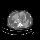
serpentine type hepatic artery pseudoaneurysms. Axial CECT arterial phase shows large poorly defined heterogenous hypodensity in right lobe of liver indicating Grade VI injury (AAST Grading). Note multiple large serpentine pseudoaneurysms in right lobe (arrow) and gross haemoperitoneum.

Post-traumatic
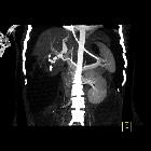
serpentine type hepatic artery pseudoaneurysms. Coronal CECT arterial phase shows the right lobe liver laceration and pseudoaneurysm.

Post-traumatic
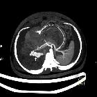
serpentine type hepatic artery pseudoaneurysms. Axial CECT portal phase image shows clearly hepatic laceration, intraparenchymal haematoma and serpentine pseudoaneurysms.

Post-traumatic
serpentine type hepatic artery pseudoaneurysms. Coronal venous phase image shows both liver and right renal inury. (HPA - Hepatic pseudoaneurysms, RK -Right kidney). Left basal lung collapse is seen.

Post-traumatic
serpentine type hepatic artery pseudoaneurysms. Axial venous phase image showing HPA. Right hepatic vein is not visualised.

Post-traumatic
serpentine type hepatic artery pseudoaneurysms. Axial MIP image shows clearly multiple irregular large serpentine (mixed fusiform-saccular) pseudoaneurysms denoted by the arrow, arising from the right hepatic artery.

Post-traumatic
serpentine type hepatic artery pseudoaneurysms. Coronal MIP image showing clearly right hepatic pseudoaneurysms (arrow).

False
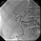
aneurysm • Hepatic artery pseudoaneurysm coiling - Ganzer Fall bei Radiopaedia

Case report:
acute pancreatitis caused by postcholecystectomic hemobilia. pseudoaneurysm.

Case report:
acute pancreatitis caused by postcholecystectomic hemobilia. right hepatic artery after embolization.

 Assoziationen und Differentialdiagnosen zu Aneurysma spurium Arteria hepatica:


