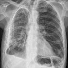
Herzwandaneurysma

Großes
Vorderwandaneurysma des linken Ventrikels mit Ausdünnung der Wand wohl nach Infarkt. Deutliche Verkalkungen auch im Röntgenbild sichtbar.


Großes
Vorderwandaneurysma des linken Ventrikels mit Ausdünnung der Wand wohl nach Infarkt. Deutliche Verkalkungen auch im Röntgenbild sichtbar.


Significant
incidental cardiac disease on thoracic CT: what the general radiologist needs to know. a, b Axial (a) and sagittal non-contrast CT image demonstrating curvilinear dystrophic calcification at the left ventricular apex in a 64-year-old female who was being evaluated for COPD. She had a history of previous myocardial infarct. b Axial non-contrast CT on bone window setting demonstrates metastatic myocardial calcification in the left ventricle in an 88-year-old woman with chronic renal impairment


Left
ventricular aneurysm • Left ventricular aneurysm - Ganzer Fall bei Radiopaedia

Left
ventricular aneurysm • Left ventricular aneurysm - Ganzer Fall bei Radiopaedia

Left
ventricular aneurysm • Calcified left ventricular aneurysm - Ganzer Fall bei Radiopaedia

Left
ventricular aneurysm • Left ventricular aneurysm - Ganzer Fall bei Radiopaedia

Left
ventricular aneurysm • Left ventricular aneurysm - Ganzer Fall bei Radiopaedia
 Assoziationen und Differentialdiagnosen zu Herzwandaneurysma:


