high malposition of the umbilical arterial catheter
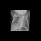
Premature
newborn after umbilical arterial and umbilical venous catheter placementCXR AP shows the tip of the umbilical arterial catheter to be in the left subclavian artery. The tip of the umbilical venous catheter is deep within the right atrium. The lungs show minimal ground-glass opacity.The diagnosis was high malposition of the umbilical arterial catheter and high malposition of the umbilical venous catheter in a patient with respiratory distress syndrome.

Premature
newborn after placement of an umbilical venous catheterAXR AP and cross-table lateral shows normal course of the umbilical venous catheter from umbilical vein to left portal vein to ductus venosus to left hepatic vein to inferior vena cava (in and cephalad on the AP view) while coursing through the liver on the lateral view with the catheter tip positioned at the junction of the inferior vena cava and right atrium. The umbilical arterial catheter has a normal course from umbilical artery to internal iliac artery to common illiac artery to aorta (in and caudad and then cephalad on the AP view) while coursing anterior to the spine on the lateral view with the catheter tip at T5. The diagnosis was appropriate position of the umbilical venous catheter and high malposition of the umbilical arterial catheter.
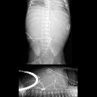
Premature
newborn after placement of an umbilical venous catheterAXR AP (above) and cross-table lateral AXR (below) shows the tip of the umbilical venous catheter to be positioned deep within the right portal vein. The tip of the umbilical arterial catheter is at T4.The diagnosis was low malposition of the umbilical venous catheter and high malposition of the umbilical arterial catheter.
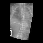
Premature
newborn after umbilical catheter placementCXR AP shows the tip of the umbilical venous catheter within the right portal vein. The tip of the umbilical arterial catheter is at T5. There is ground-glass opacity in the lungs.The diagnosis was low malposition of the umbilical venous catheter and high malposition of the umbilical arterial catheter in a patient with respiratory distress syndrome.
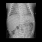
Premature
newborn after umbilical catheter placementAXR AP shows the tip of the umbilical venous catheter within the right portal vein. The tip of the umbilical arterial catheter is at T8.The diagnosis was low malposition of the umbilical venous catheter and high malposition of the umbilical arterial catheter.
high malposition of the umbilical arterial catheter
Siehe auch:


 Assoziationen und Differentialdiagnosen zu high malposition of the umbilical arterial catheter: