Infarkt Medulla oblongata

Bilateral
medial medullary infarction in association with a vertebral artery aneurysm. Axial image of diffusion-weighted MR sequence shows heart-shaped bilateral hyperintensity (arrow) in anterior and medial portion of medulla oblongata.

Bilateral
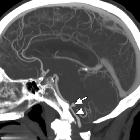
medial medullary infarction in association with a vertebral artery aneurysm. Sagittal MIP reconstruction of CT angiogram shows aneurysm (arrow) connected to right vertebral artery (arrowhead) by a narrow aneurysmal neck.

A radiologic
review of hoarse voice from anatomic and neurologic perspectives. Lateral medullary infarction. A 33-year-old woman with history of migraines presenting with acute onset vertigo, nausea, weak voice/swallow, and left extremity sensorimotor deficits after chiropractic manipulation. Diffusion-weighted MRI (a) reveals numerous acute embolic infarctions involving the bilateral cerebellar hemispheres and bilateral thalami, with notable involvement of the left PICA territory including the left lateral medulla (black arrowhead). TOF MRA MIP image (b) reveals absence of flow related enhancement within the left V4 segment and PICA (white arrowhead). Axial TOF image (c) reveals small caliber flow within the residual true lumen (white arrow), with surrounding crescentic lower signal in the false lumen. This constellation of findings is consistent with left vertebral artery dissection with showering of emboli resulting in infarction

Nicht mehr
frischer Defekt dorsolateral in der Medulla oblongata rechts mit der Klinik eines Wallenberg-Syndroms. Oben axial T2, DWI und T1 mit KM sagittal; unten axial T1 mit KM, ADC und FLAIR sagittal.
Infarkt Medulla oblongata
Siehe auch:

 Assoziationen und Differentialdiagnosen zu Infarkt Medulla oblongata:
