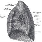
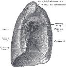
inferior pulmonary ligament


The inferior pulmonary ligament (or just the pulmonary ligament) is a normal anatomical structure that is often seen on chest x-ray and CT chest.
Gross anatomy
The inferior pulmonary ligament is a fused triangular-shaped sheet of parietal and visceral pleura that extends from the hilum to the dome of the hemidiaphragm. It extends from the mediastinum to the medial surface of the lower lobe and is extra-parenchymal to the lung. It exists to allow vascular enlargement of the hilar vessels in times of increased cardiac output.
Contents
- inferior pulmonary vein (at the apex)
- intrapulmonary lymph nodes
- bronchial veins
Radiographic features
CT
- thin, linear and predominantly midline hyperdensity that arises inferiorly to the hilum on lung windows
- most apparent at the level of the hemidiaphragm
- twice as commonly seen on the left than the right; evident in ~50% of patients
Differential diagnosis
- inferior accessory fissure
- sublobar septum
- pleural effusion

