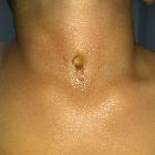
infizierte Ductus thyreoglossus Zyste

Thyroglossal
duct pathology and mimics. Infected thyroglossal duct cyst. A 23-year-old male presents with several days of fever, as well as a warm anterior neck mass. Long axis ultrasound image with color Doppler reveals a thick-walled hypoechoic structure containing low level echoes with peripheral hyperemia in the paramidline anterior neck

Thyroglossal
duct pathology and mimics. Infected thyroglossal duct cyst with abscess. a Sagittal contrast-enhanced CT image of the neck shows a thick-walled cyst adjacent to the hyoid bone (arrow). There is a communicating peripherally enhancing fluid collection along the floor of the mouth, consistent with abscess (arrowhead). Note stranding of the adjacent fat. b Sagittal post-contrast T1-weighted MR imaging of the neck demonstrates interval resolution of the abscess and underlying thyroglossal duct cyst with residual inflammatory changes. Note the relative increased thickness of the wall of the infected thyroglossal duct cyst compared to one that is not infected (see Fig. 2)
infizierte Ductus thyreoglossus Zyste
Siehe auch:

 Assoziationen und Differentialdiagnosen zu infizierte Ductus thyreoglossus Zyste:


