inflammatorischer Pseudotumor der Milz

Splenic
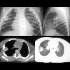
inflammatory (myofibroblastic) tumour: CT and MRI findings. Arterial-phase (b,c) images showed inhomogeneous, predominantly peripheral enhancement of the demarcated splenic mass (arrowheads).

Splenic
inflammatory (myofibroblastic) tumour: CT and MRI findings. Peripheral contrast enhancement progressed during the portal venous (d,e) phase. The ovoid splenic mass (arrowheads) appeared to be well-demarcated and measured approximately 4.5x6 cm.

Splenic
inflammatory (myofibroblastic) tumour: CT and MRI findings. Coronal (a), axial (b) and fat-suppressed (c) T2-weighted images confirmed well-demarcated ovoid lesion (arrowheads) at the upper splenic pole, with lower signal intensity peripherally compared to the spleen (*).
inflammatorischer Pseudotumor der Milz
Siehe auch:
- Raumforderungen der Milz
- inflammatorischer Pseudotumor
- rupturierter inflammatorischer Pseudotumor der Milz
und weiter:


 Assoziationen und Differentialdiagnosen zu inflammatorischer Pseudotumor der Milz: