Inflammatory myofibroblastic tumor of the lung


Inflammatory myofibroblastic tumors of the lungs are a location-specific type of inflammatory myofibroblastic tumors.
Epidemiology
They are very rare with their incidence reported at approximately 0.04-1% of all the pulmonary neoplasms . While it can affect any age group, around 25% of cases occur in those under 18 years of age. It is the most common primary mass of the lung in children.
Pathology
It is generally considered to fall within the benign spectrum of tumors.
Microscopically they are characterized by abundant inflammatory infiltrate comprising of predominantly plasma cells, lymphocytes, histiocytes, admixed with a variable proportion of fibroblasts and myofibroblasts.
On immunohistochemistry, the tumor cells can exhibit
- strong diffuse positivity with smooth muscle actin and vimentin
- negativity for cytokeratin, CD34 and S100
Subtypes
They are sometimes divided into two subtypes:
- non-invasive
- invasive: usually occurs among younger patients and may reach large sizes
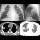
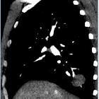
Radiographic features
CT
Typically seen as a single (can be multiple in ~5% of cases) peripheral, lobulated mass with lower lobe predominant occurrence. Calcification can occur (commoner in tumors occurring in children). Usually shows heterogeneous enhancement with contrast.
Nuclear medicine
Usually avid on FDG PET-CT.
Treatment and prognosis
Its behavior is often variable and the overall prognosis is often dependent on tumor size. Radical resection with negative margins is often the mainstay of treatment while radiation and steroids may be options in those who cannot undergo surgery. Long-term surveillance is often recommended due to local and distant recurrence as well as possible sarcomatous degeneration.
History and etymology
They were first observed in the lungs and described by Brunn in 1939.
Siehe auch:
- pulmonales Hamartom
- solitärer pulmonaler Rundherd
- inflammatorischer Pseudotumor
- pulmonary pseudotumours
- inflammatory pseudotumour IgG4 related disease
und weiter:


 Assoziationen und Differentialdiagnosen zu entzündlicher Pseudotumor der Lunge: