interkostale Lungenhernie
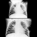


Intercostal lung hernias are defined as a protrusion of the lung beyond the confines of the thoracic cage. They are uncommon, mostly seen post trauma or thoracotomies.
Clinical presentation
Hernias which are symptomatic may cause dyspnea, chest wall pain or a visible or palpable chest bulge (most common in intercostal lung hernias). They may also be asymptomatic.
Pathology
Lung hernias are classified by their anatomic locations and the mechanism by which they arise (congenital or acquired).
Etiology
They can be either congenital or acquired in origin (classified by Morel-Lavallée in 1847):
- congenital
- acquired (most common)
- pathological
- inflammatory
- traumatic
- iatrogenic, e.g. post-thoracotomy incision
Location
- cervical: ~35%
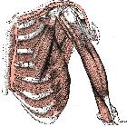
- protrusion of the lung through the anterior region of the thoracic inlet where there is a space between the scalenus anterior and sternocleidomastoid muscles
- mostly seen in elderly patients with emphysematous lungs with weak cervical fascia
- intercostal: ~70% (range 60-83%)
- result of the weakening of the thoracic wall or abnormally elevated intrathoracic pressure (e.g. weightlifters, wind instrument players)
- in post-traumatic cases, the lung herniation may occur immediately after the impact or years later
- diaphragmatic: extremely rare
Treatment and prognosis
Asymptomatic lung hernias may be managed by close observation. In symptomatic cases, immediate reduction and closure of the defect are indicated to prevent incarceration and strangulation .
Although lung hernias are rare and usually benign in nature, it is important for physicians to be aware that these entities do exist so that they are not alarmed when they are encountered. Knowledge of the benign nature of lung hernias will prevent the use of unnecessary invasive procedures and surgery.
Siehe auch:
- Musculus sternocleidomastoideus
- Lungenhernierung durch Sternotomie
- Musculi intercostales externi
- apical lung herniation
- Musculi intercostales intimi
- Musculus intercostalis internus
und weiter:


 Assoziationen und Differentialdiagnosen zu interkostale Lungenhernie: