intracanalicular acoustic schwannoma

Imaging of
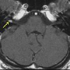
cranial nerves: a pictorial overview. Small intracanalicular vestibulocochlear schwannoma. MRI steady-state free procession (SSFP) (a) and T1-weighted post-gadolinium (b) axial images show a small hypointense lesion on steady-state sequence with avid contrast-enhancement visible in the intracanalicular segment of internal auditory canal (arrows)
Winziges
de:Schwannom des rechten de:Nervus vestibularis, inkorrekte aber verbreitete Bezeichnung: de:Akustikusneurinom, im inneren Gehörgang rechts (im Bild links, mit Pfeil markiert). Transversale T1-gewichtete de:Kernspintomographie nach Applikation von Kontrastmittel. Tumoren dieser Größe sind ausschließlich mit der Kernspintomographie nachweisbar.

Schwannom im
inneren Gehörgang links (rechts im Bild) in der MRT T1 mit Kontrastmittel.
intracanalicular acoustic schwannoma
Siehe auch:


 Assoziationen und Differentialdiagnosen zu intracanalicular acoustic schwannoma: