intraduktale papilläre Neoplasien der Mamma
Intraductal
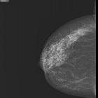
papillary neoplasm. Right MLO view: Multiple, well-defined, rounded, radio-opaque structures in the right retroareolar region with surrounding low attenuating hallow. No enlarged axillary lymph nodes.

Intraductal
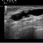
papillary neoplasm. Ultrasound: A well-defined, echogenic, solid-appearing lesion in the dilated duct. Adjacent dilated ducts are also noted.

Intraductal
papillary neoplasm. Ultrasound: Multiple echogenic, solid lesion in dilated ducts.

Intraductal
papillary neoplasm. Right CC view: Multiple, well-defined, rounded, radio-opaque structures in the right retroareolar region with surrounding low attenuating hallow. No focal calcification in the vicinity. No evidence of skin thickening or nipple retraction.French Medical Institute for Children
intraduktale papilläre Neoplasien der Mamma
Siehe auch:


 Assoziationen und Differentialdiagnosen zu intraduktale papilläre Neoplasien der Mamma:
