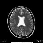
isolierte Agenesie des Septum pellucidum

Isolated
Agenesis of Septum Pellucidum. Figure 1b: Sagittal T1W image showing normal corpus callosum and neurohypophysis.

Isolated
Agenesis of Septum Pellucidum. Figure 1c: Axial T2W image showing fusion of both lateral ventricles in their bodies and more or less normal frontal horns and atria of the lateral ventricles. Rest of both cerebral hemispheres are normal.

Isolated
Agenesis of Septum Pellucidum. Figure 1d: Axial T2W image showing fusion of both lateral ventricles in their bodies. Rest of both cerebral hemispheres are normal.

 Assoziationen und Differentialdiagnosen zu isolierte Agenesie des Septum pellucidum:
