juvenile papillomatosis of the breast


Juvenile papillomatosis (JP) of the breast is a relatively common benign localized proliferative lesion in the breast.
Epidemiology
As the name implies, it is mainly seen in young women (mean age ~19-23 years ) and is unusual in women over 30 years old.
Clinical presentation
Patients present with a firm, well-defined, mobile mass often in the periphery of the breast. There is usually no nipple discharge.
Pathology
Juvenile papillomatosis is a papillary proliferation of the ductal epithelium which partly fills up smaller ducts and distends them to a degree. Gross pathology often shows a well-circumscribed mass containing multiple small cysts (<2 cm) within a dense fibrous stroma (therefore sometimes termed Swiss cheese disease by pathologists). Lesions can vary in size, usually range from 1 to 8 cm.
Radiographic features
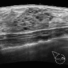
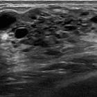
Breast ultrasound
They can appear as an ill-defined, inhomogeneous hypoechoic mass with multiple small (up to 4 mm) predominantly peripheral cysts. Microcalcifications may be seen at sonography .
Mammography
These lesions are usually negative on mammography . Occasionally mammograms may show pleomorphic or amorphous microcalcifications, an asymmetric density or a prominent intraductal pattern.
Galactography
May show a multiple irregular filling defects within the breasts.
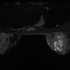
Breast MRI
Signal characteristics according to one report include:
- T1: hypointense lobulated mass
- T2: may show presence of multiple small internal cysts, best shownon this sequence and considered the most specific feature
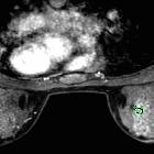
- T1 C+ (Gd): may show marked rapid enhancement
- dynamic sequence: shows a benign enhancement profile
Treatment and prognosis
Despite being a benign entity, it is considered by some to be a marker for familial breast cancer. Approximately 10% of those with papillomatosis are thought to develop breast cancer later in life .
Siehe auch:
und weiter:


 Assoziationen und Differentialdiagnosen zu Juvenile Papillomatose der Mamma:
