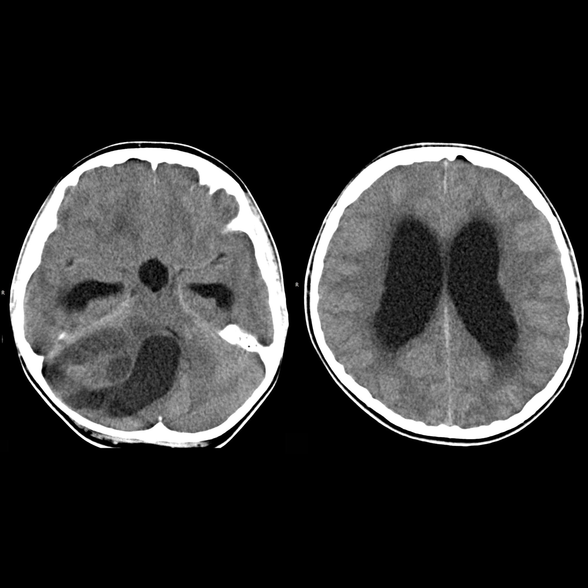
juvenile pilocytic astrocytoma of the cerebellum
Role of
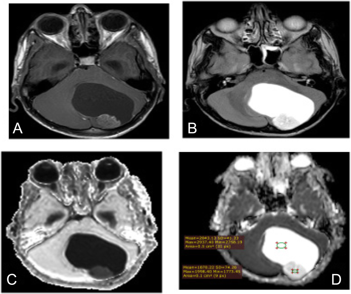
diffusion-weighted imaging in differentiation between posterior fossa brain tumors. Juvenile pilocytic astrocytoma in a 9-year-old boy who presented with repeated headache and vomiting. a T1-weighted (T1WI) shows intra-axial cystic space-occupying lesion (SOL) in the left cerebellar hemisphere. b T2-weighted (T2WI) showing hyperintense cystic posterior fossa mass with isointense solid mural nodule attached to the inner side of the cystic mass. Note the cystic components and relatively solid components. c Post-contrast T1WI axial image showing mildly enhancing solid components and faint enhanced cystic wall. d Apparent diffusion coefficient (ADC) map: the cystic component is bright while the solid component is relatively lower than the cystic component with slight diffusion restriction
juveniles zerebelläres pilozytisches Astrozytom
juvenile pilocytic astrocytoma of the cerebellum
Siehe auch:


 Assoziationen und Differentialdiagnosen zu juveniles zerebelläres pilozytisches Astrozytom: