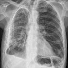
kardiale Verkalkungen

Großes
Vorderwandaneurysma des linken Ventrikels mit Ausdünnung der Wand wohl nach Infarkt. Deutliche Verkalkungen auch im Röntgenbild sichtbar.

Verkalkungen
an den Spitzen der Papillarmuskeln der Mitralklappe. Darstellung in der Computertomographie. Man erkennt auch Kalk an der Klappe selbst.

Ausgeprägte
Verkalkungen der Mitralklappe bei einer 86-jährigen im Röntgenbild.

Significant
incidental cardiac disease on thoracic CT: what the general radiologist needs to know. a, b Axial (a) and sagittal non-contrast CT image demonstrating curvilinear dystrophic calcification at the left ventricular apex in a 64-year-old female who was being evaluated for COPD. She had a history of previous myocardial infarct. b Axial non-contrast CT on bone window setting demonstrates metastatic myocardial calcification in the left ventricle in an 88-year-old woman with chronic renal impairment

Significant
incidental cardiac disease on thoracic CT: what the general radiologist needs to know. Axial CT image demonstrates chunky pericardial calcification on a non-contrast image, in the AV grooves and bilaterally in a 36-year-old male being investigated for chronic pain

Caseous
calcification as a cause of heart failure evaluated by multimodality imaging. Contrast-enhanced cardiac CT long-axis 2 chambers multiplanar reconstruction (MPR) confirms the massive calcification of the mitral annulus (arrow).


Senile
Cardiac Calcification Syndrome: A Rare Case of Extensive Calcification of Left Ventricular Papillary Muscle: (a) Multidetector computed tomography demonstrated a spindle-like extensive calcification of anterolateral papillary muscle (arrow); (b) diffuse mild stenosis with calcified plaques in proximal portion of left anterior descending coronary artery disease and mild segmental stenosis with calcified plaques in proximal portion of obtuse marginal artery (arrow).
 Assoziationen und Differentialdiagnosen zu kardiale Verkalkungen:


