Katzenkratzkrankheit

Cat-scratch
disease encephalitis. Using the convex transducer probe subtle small hypoecoic lesions were identified (yellow arrows).

Cat-scratch
disease encephalitis. After changing to a high-frequency linear probe, multiple hypoechoic liver and splenic lesions became more conspicuous (yellow arrows), consistent with hepato-splenic granulomata or micro-abscesses.

Cat-scratch
disease encephalitis. Axial DWI images of the brain showing scattered cortical altered diffusion (blue arrows), probably related to cerebral vasogenic oedema.

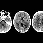
Cat-scratch
disease encephalitis. Unenhanced brain CT showed no relevant abnormality.
Cat-scratch
disease encephalitis. Liver MRI confirmed the presence of multiple T2-hyperintense lesions showing restricted diffusion (red arrows) compatible with granulomata or micro-abscesses.

Cat-scratch
disease encephalitis. One week after the acute onset of CSD encephalitis, abdominal sonography was performed. The hypoechoic lesions have increased in size, possibly related to post-treatment changes.

Cat-scratch
disease encephalitis. Sonography revealed a hypoechoic rounded lesion (yellow arrows) with a vascular hilum (blue arrow), with an adjacent subcutaneous collection (red arrows), consistent with supurative inflamatory lymphadenopathy.Prat-Matifoll J.A, Department of Radiology, Vall Hebron Hospital

Imaging of
parotid anomalies in infants and children. Cat scratch disease. Left, intraparotid necrotic lymphadenitis: ultrasonography (a); axial T1-weighted image, after intravenous administration of gadolinium chelate (b)


 Assoziationen und Differentialdiagnosen zu Katzenkratzkrankheit: