Kongenitale Skoliose

School ager
with a posterior mediastinal mass. CXR AP shows a round soft tissue density in the superior and medial aspect of the right hemithorax which was found on cross sectional imaging to be in the posterior mediastinum, along with multiple hemivertebra segmentation anomalies of the lower cervical and upper thoracic spine causing a curvature of the upper thoracic spine convex to the right.The diagnosis was congenital scoliosis secondary to hemivertebra in a patient with a neurenteric cyst.

Infant with
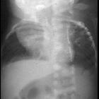
respiratory distress. CXR AP shows multiple left-sided anterior and multiple right-sided posterior rib fusions and a mild curvature of the spine convex left resulting in a small thorax.The diagnosis was congenital scoliosis secondary to rib fusion causing pulmonary hypoplasia.

Infant with a
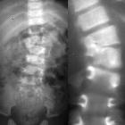
curvature to the spine. AP radiograph of the lumbar spine (left) demonstrates a curvature of the thoracolumbar spine convex to the left caused by a fusion of the right sided pedicles of the L1 and L2 vertebral bodies which is better demonstrated on the tomographic image (right).The diagnosis was congenital scoliosis due to a congenital bar.

Newborn with
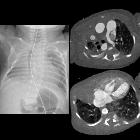
complex congenital heart disease on prenatal US. CXR AP (left) shows the right hemithorax to be smaller than the left hemithorax and there is opacity in the right upper and middle lobes and an aerated right lower lobe. There are multiple hemivertebrae and a butterfly vertebra in the thoracic spine causing a scoliosis convex left. Axial CT with contrast of the chest (above right) shows the right pulmonary artery to be absent along with the right upper lobe. Axial CT (below right) shows absence of the right middle lobe of the lung but the right lower lobe of the lung is present and aerated via a small bronchus off of the trachea (not shown) and perfused by collateral vessels arising from the 7 o’clock position off of the aorta.The diagnosis was right pulmonary agenesis with the hypoplastic right lower lobe receiving its arterial supply from collaterals off of the aorta in a patient who has multiple vertebral body anomalies resulting in congenital scoliosis.

Newborn with
absent anus and stool coming out of the vagina. CXR AP (left) shows a hemivertebra at L1 causing spinal curvature convex left. Transverse US of the pelvis (above right) shows in the midline anteriorly an anechoic fluid-filled bladder with a round echogenic stool-filled rectum posterior to it while a transverse US of the perineum (below right) shows a very short distance between the calipers superiorly on the skin and inferiorly on the anterior wall of the rectum. The diagnosis was low anorectal malformation and congenital scoliosis.

Toddler with
short stature. AP and lateral radiographs of the spine show multiple vertebral body anomalies throughout the spine including butterfly vertebrae, hemivertebrae, fused vertebrae and hypoplastic vertebrae. There are also multiple levels of rib fusion present posteriorly.The diagnosis was spondylothoracic dysostosis with the multiple vertebral body anomalies resulting in congenital scoliosis.
Kongenitale Skoliose
Siehe auch:
und weiter:

 Assoziationen und Differentialdiagnosen zu Kongenitale Skoliose:

