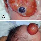
kongenitales Teratom der Orbita

Orbitales,
retrobulbäres Teratom bei einem 2 Monate alten Kind, somit kongenital: Oben T2 axial, T1 axial, T2 koronar; unten T1 KM 3 Ebenen

Congenital
orbital teratoma: a case report with preservation of the globe and 18 years of follow-up. a CT of the orbits shows a large heterogeneous mass, filling the entire orbit. b T2-weighted MRI shows the tumor to be hyperintense to fat and extraocular muscles

Congenital
orbital teratoma in fetal MRI. Enlargement of the left globe, with a mass at its posterior aspect extending through the optic canal.

Congenital
orbital teratoma in fetal MRI. Mass at posterior aspect left eye globe. No intracranial extension.

Congenital
orbital teratoma in fetal MRI. Axial CT reveals enlargement of the left orbit containing a predominantly cystic mass. Minor associated calcification in the posterior globe. No identifiable lens or anterior chamber.

Congenital
orbital teratoma in fetal MRI. Axial T2W: left orbital cystic mass with well defined margins and a solid component at its posterior aspect. No globe separately identified.

Congenital
orbital teratoma in fetal MRI. Sagittal T1W: solid component noted at the posterior aspect of the cystic orbital lesion. No intracranial extension. Extraconal optic nerve projecting posteriorly from the mass. No globe separately identified.
kongenitales Teratom der Orbita
Siehe auch:
und weiter:

 Assoziationen und Differentialdiagnosen zu kongenitales Teratom der Orbita:
