Leiomyosarkom der Bartholinschen Drüsen

Vulvar
leiomyosarcoma in Bartholin"s gland. Right vulvar mass of intermediate signal intensity.

Vulvar
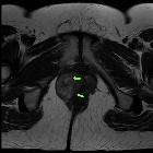
leiomyosarcoma in Bartholin"s gland. Axial T2 TSE. Relative isointense mass centred in the right vulva, replacing the labia majora and showing central areas of hyperintensity (green arrows).

Vulvar
leiomyosarcoma in Bartholin"s gland. Coronal T2 TSE. The mass is located in Bartholin"s gland area and extends to the right elevator muscle of anus (yellow arrows), without showing invasion.

Vulvar
leiomyosarcoma in Bartholin"s gland. Sagittal T2 TSE. The lesion is superior to the perineal membrane (orange arrow).

Vulvar
leiomyosarcoma in Bartholin"s gland. Early phase: the mass depicts heterogeneous, mainly peripheral enhancement.

Vulvar
leiomyosarcoma in Bartholin"s gland. Delayed phase: progressive and heterogeneous fill-in of the lesion.

Vulvar
leiomyosarcoma in Bartholin"s gland. Heterogeneous, high-signal-intensity mass (asterisk) on high b-value DWI.

Vulvar
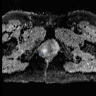
leiomyosarcoma in Bartholin"s gland. Heterogeneous, low-signal-intensity mass (asterisk), confirming the restricted diffusion on ADC map.
Leiomyosarkom der Bartholinschen Drüsen
Tumoren der Bartholinschen Drüsen Radiopaedia • CC-by-nc-sa 3.0 • de
Bartholin gland tumors represent neoplasms of the Bartholin glands.
They include:
- squamous cell carcinoma of the Bartholin gland: tends to be the most common histological subtype
- adenocarcinoma of the Bartholin gland
- adenoid cystic carcinoma of the Bartholin gland
Siehe auch:

 Assoziationen und Differentialdiagnosen zu Leiomyosarkom der Bartholinschen Drüsen:
