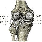
Ligamentum meniscofemorale anterius

Ligamentum
meniscofemorale anterius (Humphry) and ligamentum meniscofemorale posterius (Wrisberg) in MRI. There is effusion of blood in the joint cavity from a trauma wich did not affect the posterior ligaments.

Left
knee-joint from behind, showing interior ligaments including the meniscofemoral ligaments Humphry and Wrisberg.
Ligamentum meniscofemorale anterius
Ligamentum meniscofemorale Radiopaedia • CC-by-nc-sa 3.0 • de
The meniscofemoral ligament (MFL) arises from the posterior horn of the lateral meniscus and passes to attach to the lateral aspect of the medial femoral condyle. It splits into two bands at the posterior cruciate ligament (PCL), which are named in relation to the PCL:
- anterior meniscofemoral ligament (ligament of Humphrey)
- posterior meniscofemoral ligament (ligament of Wrisberg)
A handy mnemonic to recall the relationship is here.
Variant anatomy
The MFL is variably described as either possessing one band (~35%) or as described above, possessing two bands (~65%) .
Approximately 80% (range 65%-100%) of people will have at least one MFL with the posterior MFL (50-70%) more commonly present than the anterior MFL (10%-25%). 20-30% of people will have both anterior and posterior MFLs .
Related pathology
- tear of the meniscofemoral ligament: Wrisberg rip
- on sagittal MR images, the MFL may mimic an intra-articular loose body or meniscal fragment
Siehe auch:
- Ligamentum meniscofemorale
- Ligamentum meniscofemorale posterius
- Meniskusriss
- popliteomeniskale Faszikel
- Ligamentum meniscotibiale
- accessory ligaments of the knee
und weiter:

 Assoziationen und Differentialdiagnosen zu Ligamentum meniscofemorale anterius:

