Lipidpneumonie


Lipoid pneumonia is a form of pneumonia associated with oily or lipid components within the pneumonitis component.
This can either result from
- aspiration of oily substances (exogenous lipoid pneumonia) or
- endogenous accumulation of lipid substances in the alveoli (endogenous lipoid pneumonia).
Clinical presentation
Most patients are asymptomatic and often discovered incidentally.
Pathology
Lipid-laden macrophages are often seen in histological samples following transthoracic needle biopsy. With exogenous forms, inhaled lipid content (e.g. from aspiration) is phagocytosed by macrophages which fill alveoli. A subsequent acute +/- chronic pneumonitis results.
Macroscopically the affected regions often have a yellowish or golden hue, which is thought to be produced by the liberation of lipid material from alveolar pneumocytes secondary to the inflammatory reaction.
Risk factors
- aspiration risk
- neuromuscular disorders
- esophageal abnormalities
- cleft palate
Associations
The endogenous type can be seen in association with lung cancer .
Case reports are emerging in patients who use e-cigarettes (vaping) .
Complications
A fibrotic component can develop in chronic cases.
Other possible complications include:
- superinfection by non-tuberculous mycobacteria
- respiratory insufficiency
- cor-pulmonale
- hypercalcemia
Radiographic features
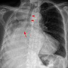
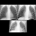
Plain radiograph
- can be variable
- radiological spectrum with consolidation to an irregular mass-like lesion to a reticulonodular pattern
CT
- characteristically show low attenuation within the consolidated areas (low attenuation consolidation) of ~ -100 HU reflecting a fat content (at times the attenuation value may be less i.e. around -30 HU and higher than that of subcutaneous fat )
- consolidation may have a predilection for the dependent portions of the lungs
- associated ossific foci may be present within the affected region
- a crazy paving pattern may also be seen
MRI
Not part of routine evaluation. Signal characteristics may reflect fat/paraffin content. usually:
- T1: high to intermediate signal
- T2: low to intermediate signal
Treatment and prognosis
Serial radiographs showing stability may be enough in asymptomatic patients with no background history. A biopsy can be performed in some of the cases to ensure the benign nature of the lesion, especially if changes are lipid-poor and imaging features persistent.
The mainstay of management in exogenous types is control and cessation of offending agent(s).
Siehe auch:
- Lungenkarzinom
- crazy paving-Muster
- Gaumenspalte
- exogene Lipidpneumonie
- chronische exogene Lipidpneumonie
- endogene Lipidpneumonie
und weiter:


 Assoziationen und Differentialdiagnosen zu Lipidpneumonie: