liposclerosing myxofibrous tumour (LSMFT)
Liposclerosing myxofibrous tumors (LSMFT), also known as polymorphic fibro-osseous lesions of bone, are rare benign fibro-osseous lesions that have a predilection for the intertrochanteric region of the femur.
Clinical presentation
It is slightly more common in males with mean age of 30-40 years. It can be discovered incidentally but mostly patients have vague longstanding pain. About 10% of patients present acutely with pathologic fracture.
Pathology
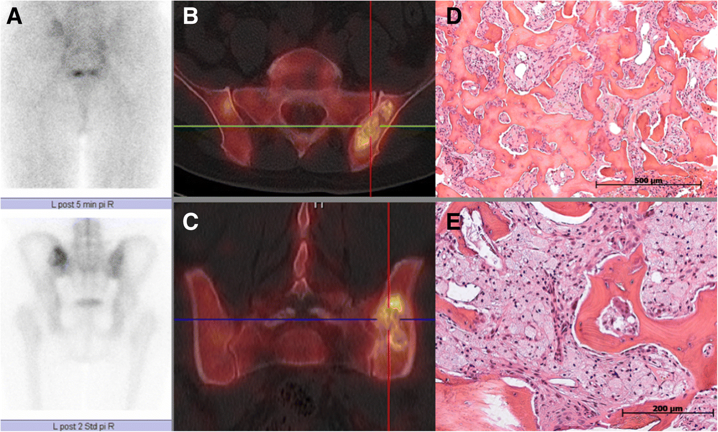
The tumor comprises of the wide mixture of tissues of lipomatous, fibroxanthomatous, myxomatous, and myxofibromatous components inclusive.
Location
Tends to have a striking predilection for the intertrochanteric region of the femur (80-90%) .
Radiographic features
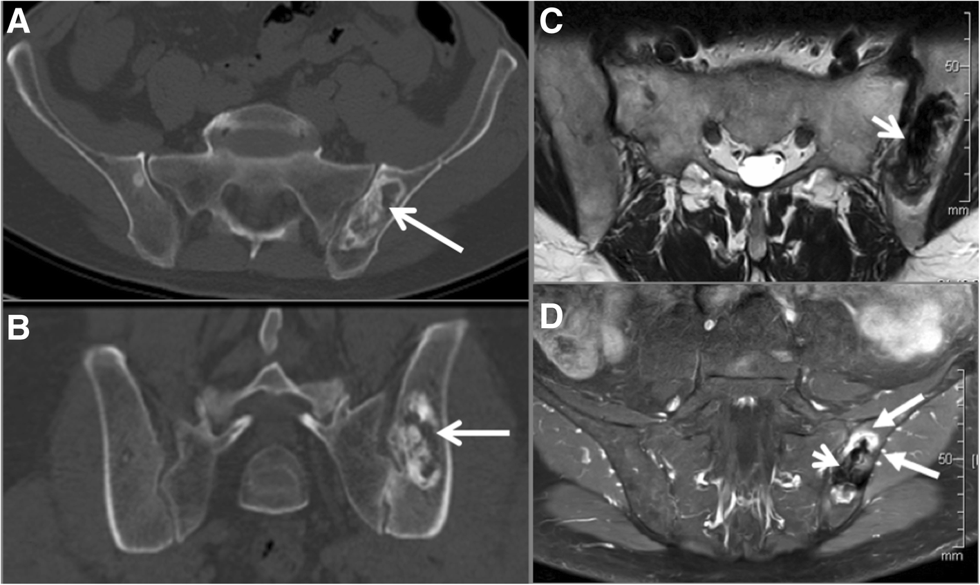
Plain radiograph/CT
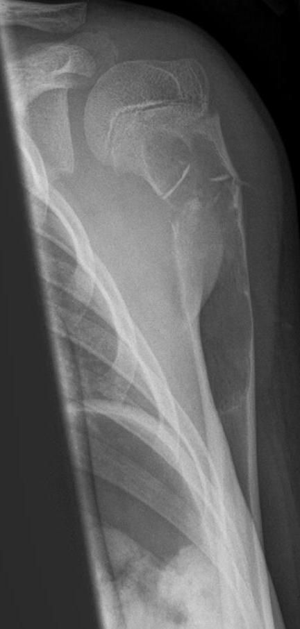
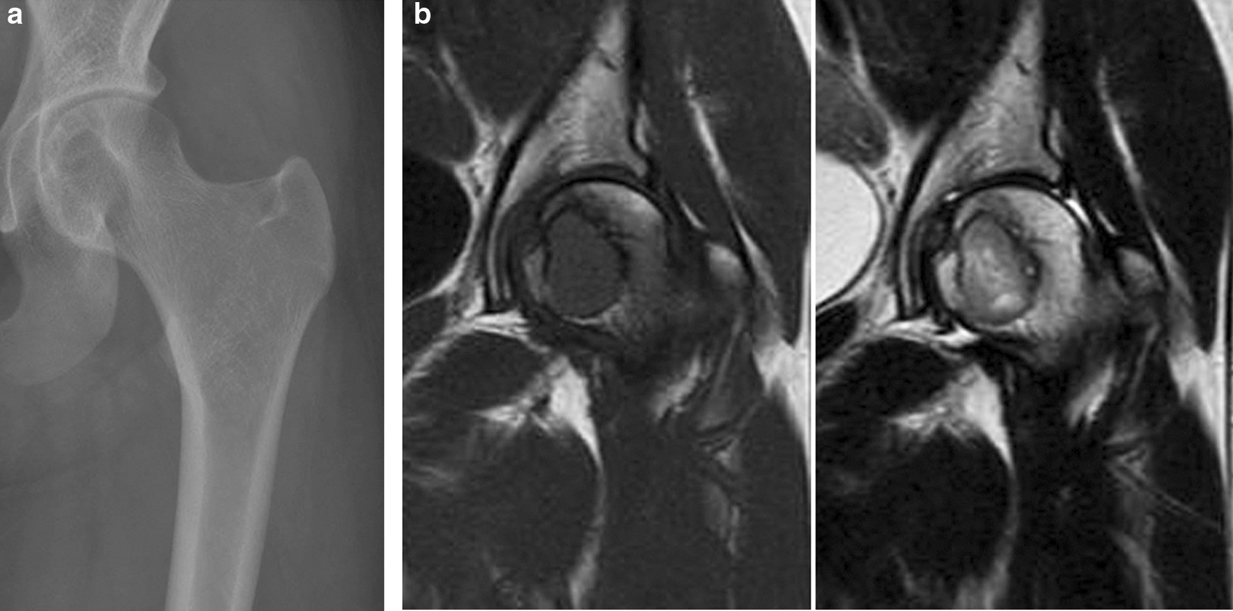
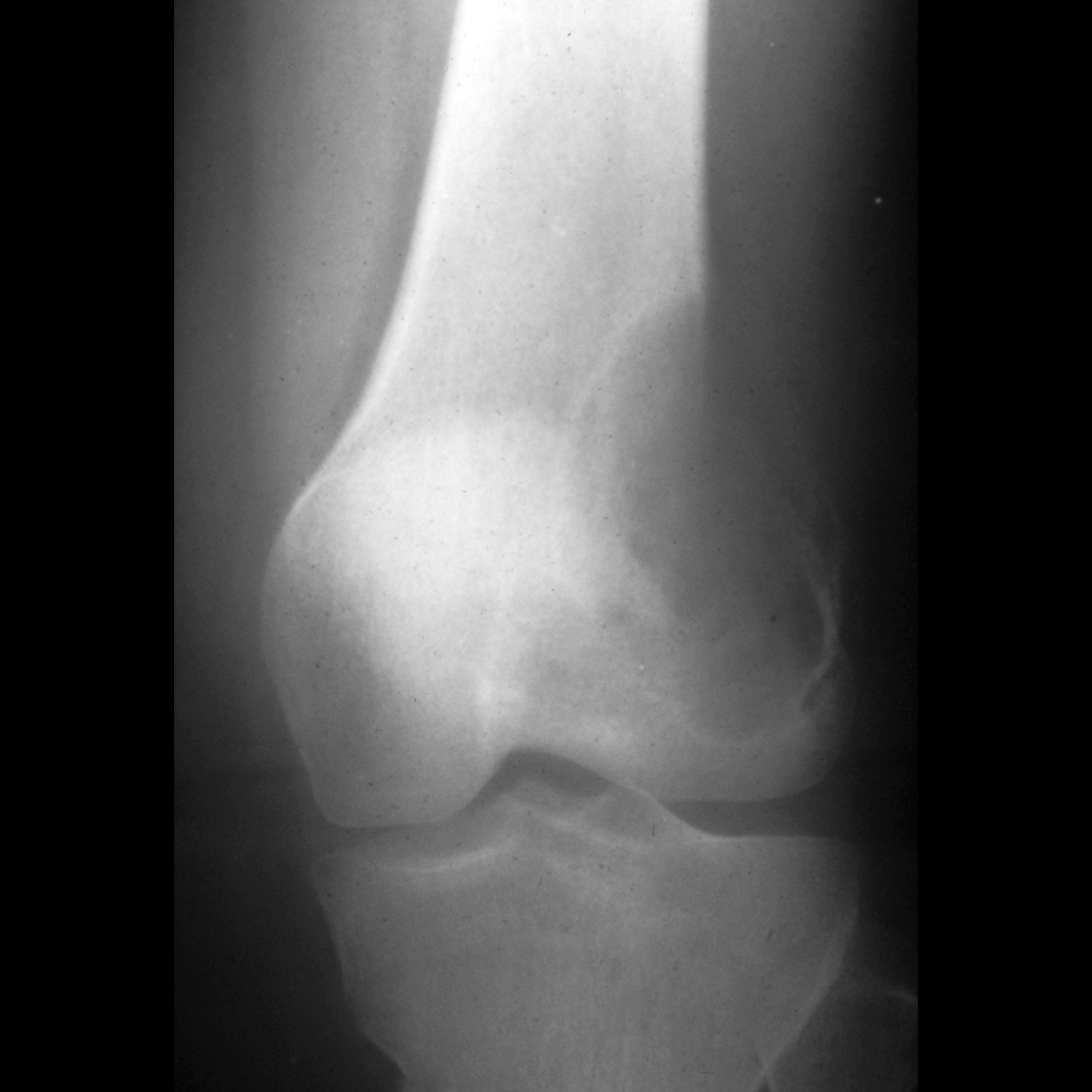
- geographic lucent lesion usually centered in intertrochanteric region of the proximal femur
- sclerotic margin
- mildly expansile
- multilocular
- matrix calcification in ~70% of cases
- fat density component
MRI
Despite its name, distinct fatty components are not seen
- T1: relatively homogeneous and isointense to skeletal muscle
- T2: moderately heterogeneous with areas of high signal due to myxoid component
Nuclear medicine
Scintigraphy can show mild focal uptake with Tc-99m pertechnetate .
Treatment and prognosis
- incidentally detected asymptomatic lesions: no treatment or intervention
- symptomatic lesions: are commonly managed with bone curettage, bone grafting, and fixation
- pathological fracture: uncommon ~10%; proximal femoral lesions may require arthroplasty
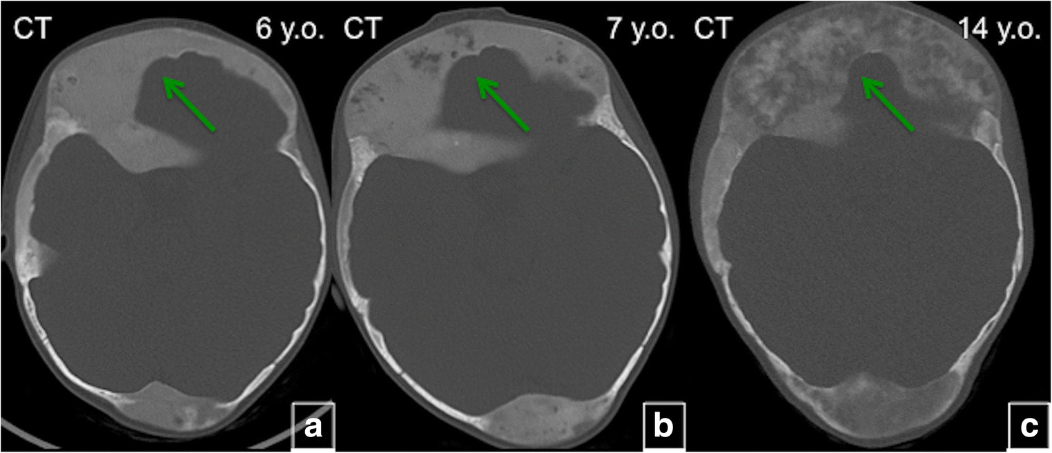
- malignant transformation: rare but documented 10-15%; transformation into osteosarcoma is the most common
Due to this potential malignant transformation, lesions need follow-up imaging preferably by MRI. Symptomatic lesions or those with interval change require surgical resection .
Differential diagnosis
On radiographs consider:
- fibrous dysplasia: it is challenging to differentiate between both by imaging. Fibrous dysplasia may show less sclerosis by radiography, more uptake by scintigraphy and intermediate or low signal intensity by fluid sensitive MRI sequences
- intraosseous lipoma: LSMFT does not usually show macroscopic fat on CT or MRI as lipomatous component is usually too small and mixed with other more prominent myxofibrous or fibro-osseous tissue
- aneurysmal bone cyst (ABC): usually more expansile
Siehe auch:
- Fibröse Dysplasie
- intraossäres Lipom
- Aneurysmatische Knochenzyste
- benigne Osteolysen
- Tumoren proximales Femur
- lytische Läsion im proximalen Femur
- Knochentumoren des Femur
und weiter:


 Assoziationen und Differentialdiagnosen zu liposklerosierender myxofibroider Tumor: