low malposition of the umbilical venous catheter

Premature
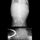
newborn after placement of an umbilical venous catheterAXR AP (above) and cross-table lateral AXR (below) shows the tip of the umbilical venous catheter to be positioned deep within the right portal vein. The tip of the umbilical arterial catheter is at T4.The diagnosis was low malposition of the umbilical venous catheter and high malposition of the umbilical arterial catheter.

Premature
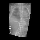
newborn after umbilical catheter placementCXR AP shows the tip of the umbilical venous catheter within the right portal vein. The tip of the umbilical arterial catheter is at T5. There is ground-glass opacity in the lungs.The diagnosis was low malposition of the umbilical venous catheter and high malposition of the umbilical arterial catheter in a patient with respiratory distress syndrome.

Premature
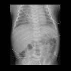
newborn after umbilical catheter placementAXR AP shows the tip of the umbilical venous catheter within the right portal vein. The tip of the umbilical arterial catheter is at T8.The diagnosis was low malposition of the umbilical venous catheter and high malposition of the umbilical arterial catheter.

Premature
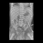
newborn after umbilical venous catheter placementCXR AP shows the tip of the umbilical venous catheter projecting over the left portal vein. The tip of the umbilical arterial catheter projects at T6. There is hazy ground glass opacity in the lungs.The diagnosis was low malposition of the umbilical venous catheter and appropriate position of the umbilical arterial catheter in a patient with respiratory distress syndrome.

Premature
newborn after umbilical venous catheter placementAXR AP shows the tip of the umbilical venous catheter curling back upon itself within the umbilical vein. The tip of the umbilical arterial catheter projects at T6. The proximal small bowel is massively dilated.The diagnosis was low malposition of the umbilical venous catheter and appropriate position of the umbilical arterial catheter in a patient with jejunal atresia.

Premature
newborn after PICC placementAXR AP and lateral shows a right lower extremity PICC with its tip within the inferior vena cava anterior to the spine at the level of T11. The tip of the umbilical venous catheter is within the ductus venosus.The diagnosis was appropriate position of the PICC and low malposition of the umbilical venous catheter.

Premature
newborn after PICC placementAXR AP shows a right lower extremity PICC which is curled back upon itself and whose tip projects over the right femoral vein. The tip of the umbilical venous catheter projects within the umbilical vein.The diagnosis was low malposition of the PICC and low malposition of the umbilical venous catheter.

Premature
newborn after PICC placementAXR AP shows a left lower extremity PICC which courses across the midline and down the right common iliac vein. The tip of the umbilical venous catheter projects over the umbilical vein. The tip of the umbilical arterial catheter projects at T6.The diagnosis was low malposition of the PICC and low malposition of the umbilical venous catheter and appropriate position of the umbilical arterial catheter.

Newborn with
abnormal prenatal echocardiogram after PICC placementAXR AP shows a left lower extremity PICC and a right femoral venous catheter both of whose tips project over a left-sided inferior vena cava. An umbilical venous catheter tip projects over the ductus venosus. An umbilical arterial catheter tip projects at the level of T9. Nasogastric tube tip projects over the stomach in the right upper quadrant. Feeding tube tip projects transpylorically over the duodenal bulb. The cardiac apex is in the right chest.The diagnosis was appropriate position of the PICC and low malposition of the umbilical venous catheter in a patient with situs inversus.
low malposition of the umbilical venous catheter

