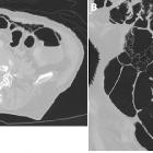
luftgefüllte Zysten am Darm

Pneumatosis
cystoides intestinalis. Follow-up axial MDCT (A) and coronal reconstruction (B) showed multiple rounded air-filled lesions occupying the anterior part of the mesentery and resolution of the pneumoperitoneum proving the imaging diagnosis of pneumatosis cystoides.
luftgefüllte Zysten am Darm
Siehe auch:

 Assoziationen und Differentialdiagnosen zu luftgefüllte Zysten am Darm: