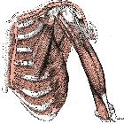
Lungenhernierung
Transkostale
Lungenhernie zwischen 9. und 10. Rippe rechts bei erweitertem Abstand zwischen diesen, mutmaßlich als Restzustand eines älteren Traumas (anamnestisch nicht zu klären).
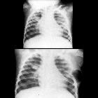
Infant who
had undergone emergency thoracotomy for cardiac massage during a cardiac arrest 6 months before and now has a lump under the left arm. CXR AP in inspiration (above) shows the left 5th intercostal space at the side of the previous thoracotomy to be deformed. CXR AP in expiration (below) shows interval development of a cystic structure in the soft tissues of the left chest wall.The diagnosis was pulmonary herniation through the left 5th intercostal space secondary to incorrectly repaired thoracotomy incision.

Traumatic
lung hernia: multidetector CT findings and relevance. Additionally, a ventral portion of the left inferior pulmonary lobe was seen herniated (arrowhead) through the 8th intercostal space. Note extrathoracic emphysema (+); nondependent pneumothorax (*).

Lung hernia
• Intercostal lung herniation - Ganzer Fall bei Radiopaedia

Lung hernia
• Intercostal lung hernia - Ganzer Fall bei Radiopaedia

Lung hernia
• Intercostal lung hernia - Ganzer Fall bei Radiopaedia

Lung hernia
• Intercostal lung herniation - Ganzer Fall bei Radiopaedia

Lung hernia
• Acquired intercostal lung hernia complicated by pneumonia - Ganzer Fall bei Radiopaedia

Lung hernia
• Intercostal lung hernia - Ganzer Fall bei Radiopaedia

Toddler with
progressive respiratory distress and no history of foreign body ingestion. CXR AP and lateral show overinflation of the right upper lobe that is herniating across the midline.

Lung
herniation post-removal of thoracostomy tube. a A soft reducible mass, prominent on coughing, valselva noted at the site of previous intercostals drainage with overlying sutures. b Chest radiograph (A-P) view showing the presence of a tongue of herniated lung tissue at the right base overlying the herniated tissue. c CT chest showing the cicatricial collapse of the right lung, tractional bronchiectatic changes in the left lung with cavities in bilateral lungs, and presence of aspergilloma in the left apical region with features of pulmonary arterial hypertension. Pleural dehiscence with lung herniation is seen in the right lung


 Assoziationen und Differentialdiagnosen zu Lungenhernierung: