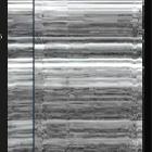
M-mode (ultrasound)

Often utilized for its excellent axial and temporal resolution of structures, M-mode (or motion mode) is a form of ultrasonography in which a single scan line is emitted, received, and displayed graphically. An M-mode recording is conventionally displayed with the abscissa representing time and the ordinate distance from the transducer, the latter derived from the time delay from echo emission to reflection and detection .
A single piezoelectric crystal is recorded and displayed graphically of which represents the acoustic impedance or density of the material encountered. These signals are subsequently displayed as dots, the brightness of which is proportional to the amplitude of reflected waves .
Physics
An alternating current directed through one of the crystals in the ultrasound transducer footprint results (via the piezoelectric effect) in its structural deformation, which results in conduction of compression-rarefaction waves through an adjacent conducting medium. If these transmitted pulses encounter an interface between two structures with a sufficiently disparate acoustic impedance, reflection may occur, which may return to the ultrasound transducer and excite the crystal.
As the speed of sound is relatively constant in soft tissues, roughly 1540 meters/second, the distance at which the transmitted pulse encountered the structure in question may be inferred based on the transit time. The distance from the transducer will be graphed on the vertical (y-axis, ordinate) plane of the rendered output, whereas the time elapsed during recording is represented by the horizontal plane (x-axis, abscissa).
The high sampling frequency, with >90% of the cycle time spent in "receive" mode and pulse transmission at a rate >1000/second, M-mode provides not only excellent temporal resolution but superior axial resolution to B-mode (i.e. the ability to discern non-contiguous structures in a vertical plane).
Clinical use
M-mode is utilized for its aforementioned superlatives; the temporal resolution is most useful in delineating the path of structures moving at a high velocity, as well as their timing (e.g. in relation to the cardiac cycle). The axial resolution is also vital in echocardiography, as it allows the resolution of delicate cardiac structures (e.g. valve leaflets) which are difficult to resolve, especially with transthoracic echocardiography. Specific examples include;
- M-mode in echocardiography
- mitral valve excursion
- axial resolution allows for the vertical distance between the maximal early excursion of the anterior leaflet of the mitral valve and the interventricular septum
- this is known as E-point septal separation (EPSS), and may be used to estimate ejection fraction
- temporal resolution allows for the timing of the early and late diastolic excursions (the E and A waves, respectively) to be related to the ECG
- right ventricular diastolic collapse, a specific sign for the presence of tamponade physiology, may be identified and related to the cardiac cycle based on the mitral valve leaflet motion
- presence or absence of systolic anterior motion (SAM) of the mitral valve with anterior leaflet prolapse into the left ventricular outflow tract
- axial resolution allows for the vertical distance between the maximal early excursion of the anterior leaflet of the mitral valve and the interventricular septum
- excursion of the lateral tricuspid annulus during systole
- commonly referred to as tricuspid annular plane systolic excursion (TAPSE), an index of right ventricular function
- mitral valve excursion
- M-mode in lung ultrasonography
- may be used to record the presence or absence of lung sliding if one is unable to store cine-loops
- differentiation of a fluid collection, such as a pleural effusion, from intraparenchymal lung pathology
- fluid dynamics dictate fluids are non-compressible, have a fixed volume, but (unlike solids) do not have a fixed shape
- respiratory maneuvers will change the shape of the pleural cavity surrounding the fluid, resulting in subtle changes which may be documented in m-mode
- pleural location identified by the quad sign
- the sinusoid sign is only present in a free-flowing fluid
Siehe auch:

 Assoziationen und Differentialdiagnosen zu M-Mode Sonographie:
