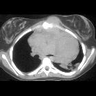
mediastinal Hodgkin's lymphoma

A diagnostic
approach to the mediastinal masses. Conventional radiograph can provide information pertaining to the size, anatomical location and density of a central mass. Right: Frontal chest radiograph shows a sharply defined area of increased opacity with a loss of the cardiac silhouette at the border of the right side of the heart (*). Contrast-enhanced CT scan reveals a thin-walled water-attenuation lesion (*) in the right cardiophrenic angle (pericardial cyst). Middle: Lateral chest radiograph and contrast-enhanced CT scan show a unilocular, well-defined and homogeneously hypodense mass in the anterior mediastinum with peripheral calcification (open arrows) (thymic cyst). Left: Chest radiograph shows the aortopulmonary window with an abnormal convex border (arrow). Contrast-enhanced CT scan demonstrates a multilobulated mass in the anterior mediastinum (arrow), which accounts for the distortion of the AP window (nodular sclerosis Hodgkin disease)
mediastinal Hodgkin's lymphoma
Siehe auch:


 Assoziationen und Differentialdiagnosen zu mediastinal Hodgkin's lymphoma: