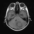
Meningeom am Tentorium

Magnetic
resonance imaging of pineal region tumours. Tentorial meningioma in a 45-year-old woman with papilloedema. A meningioma (arrow) is seen involving the tentorium which extends into the pineal recess. The lesion is isointense to grey matter on axial T2-weighted image (a) and sagittal T1-weighted image (b) and shows prominent contrast enhancement on coronal T1-weighted image (c)

Meningeom am
Tentorium mit dural tail: Oben axial T2 und T1+KM, unten coronar und sagittal T1+KM
Meningeom am Tentorium
Siehe auch:


 Assoziationen und Differentialdiagnosen zu Meningeom am Tentorium: