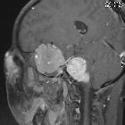
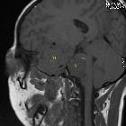
Meningeome bei Neurofibromatose Typ 2

Neurofibromatosis
type 2: proptosis as the initial presentation. Note the intense enhancement of schwannomas (s). Meningioma (m) also shows avid enhancement but slightly less as compared to schwannoma. Meningioma encased left internal carotid artery (thick arrow) and middle cerebral artery (thin arrow).

Neurofibromatosis
type 2: proptosis as the initial presentation. T2WI showing heterogenously hyperintense bilateral CPA masses (s) causing widening of internal acoustic meatuses with intracanalicular extension(*). Meningioma (m) is isointense on T2 with intraorbital extension (e).

Neurofibromatosis
type 2: proptosis as the initial presentation. On GRE, blooming black dots in schwannoma (encircled) suggest microscopic haemorrhages.

Neurofibromatosis
type 2: proptosis as the initial presentation. Intense enhancement of bilateral CPA schwannomas (s) and left sphenoid wing meningioma (m). Note meningioma extending to intraorbital extraconal space (e).

Neurofibromatosis
type 2: proptosis as the initial presentation. Enhancement of meningioma is slightly less than schwannoma.

Neurofibromatosis
type 2: proptosis as the initial presentation. Note the intraorbital extension (e) of meningioma (m).

Neurofibromatosis
type 2: proptosis as the initial presentation. m = meningioma, s = vestibular schwannoma.

Neurofibromatosis
type 2: proptosis as the initial presentation. Left sphenoid wing meningioma (m) with intraorbital extension (e).

Neurofibromatosis
type 2: proptosis as the initial presentation. Lateral rectus muscle is not distinct. Optic nerve (arrow) appears free. Meningioma possibly extends (e) through superior orbital fissure.

Neurofibromatosis
type 2: proptosis as the initial presentation. Lateral rectus muscle is not distinct. Meningioma (e) possibly extends through superior orbital fissure. It is isointense on T2WI.

Neurofibromatosis
type 2: proptosis as the initial presentation. Left sphenoid wing meningioma encasing left internal carotid artery (thick arrow) and middle cerebral artery (thin arrow) with intraorbital extension (e).

Teenager with
headache. Coronal T1 MRI with contrast of the brain shows a round, solid, homogeneously enhancing intracranial lesion above the left orbit that is in continuity with the meninges and which appears to have a dural tail. The patient also had multiple schwannomas.The diagnosis was a meningioma in a patient with neurofibromatosis Type 2.

 Assoziationen und Differentialdiagnosen zu Meningeome bei Neurofibromatose Typ 2:

