meningioangiomatosis

Multicystic
meningioangiomatosis. Axial brain magnetic resonance imaging (MRI) findings. A multicystic mass (arrow) with low signal intensity on T1-weighted images (A) and high signal intensity on T2-weighted images (B) is seen in the right temporal lobe. The cystic component was isointense with the cerebrospinal fluid on all sequences.
Three cases
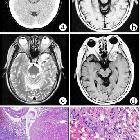
of sporadic meningioangiomatosis with different imaging appearances: case report and review of the literature. Solid meningioangiomatosis. (a) CT scan showed an irregular mixed high-density mass in the left middle cranial fossa. (b) On T1WI, the lesion demonstrated low and equal signal intensity. (c) On T2WI, the lesion showed high signal intensity with a multiple flow void effect. (d) On post-contrast MRI, the lesion showed significant and homogeneous enhancement. (e, f) Microphotography of specimens showed extensive fibroblastic proliferation and an increased number of vessels surrounded by meningothelial cells.
Three cases
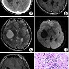
of sporadic meningioangiomatosis with different imaging appearances: case report and review of the literature. Cystic meningioangiomatosis with a cystic-mural nodule pattern. (a) CT scan showed a cystic-mural nodule lesion in the right insular lobe. (b-d) In non-enhanced MRI, the cystic content demonstrated similar signal intensity as cerebrospinal fluid (CSF), while the mural nodule demonstrated iso-signal intensity on T1WI, T2WI, and DWI. (e) On post-contrast MRI, the mural nodule demonstrated significant enhancement, while the cystic wall and content showed no enhancement. (f) Pathological examination showed perivascular spindle-cell proliferation.

Three cases
of sporadic meningioangiomatosis with different imaging appearances: case report and review of the literature. Cystic meningioangiomatosis with a multiple microcystic pattern. (a, b) DWI and T2WI demonstrated a low signal intensity nodule in the left parietal cortex and multiple small cysts surrounding it. (c) On post-contrast MRI, the nodule was remarkably enhanced. (d) Microscopically, fibroblast-like spindle cells were arranged in a spiral shape around multiple vessels, and the cortical neurons were entrapped within the lesion.
Meningioangiomatosis is a rare meningovascular hamartomatous plaque like or mass like cortical lesions extending to the overlying lepomeninges (crosses the boundary between intra and extra axial lesions). Given its frequent cortical location, often patients present with seizures. It can be sporadic or associated with neurofibromatosis type 2.
Radiographic features
These are slow growing solitary or multiple cortical lesions with nodular or gyriform configuration, variable calcification, and cystic degeneration. These show minimal or no contrast enhancement.
Siehe auch:


 Assoziationen und Differentialdiagnosen zu meningioangiomatosis: