Morgagni-Hernie mit Hernierung des linken Leberlappens
Unusual
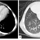
association of parvo-virus with Morgagni hernia, mistaken for patch of consolidation. Axial mediastinal (A) and lung (B) window showed a homogenous soft tissue density in RT cardio-phrenic angle abutting and displacing the pericardium and RT ventricle with clear interface. Sharp delineation with lung parenchyma is noted.

Unusual
association of parvo-virus with Morgagni hernia, mistaken for patch of consolidation. Chest X-ray PA view shows radio-opacity involving the RT cardio-phrenic angle partially silhouetting the RT cardiac border. The radio-opacity showed sharp interface with adjacent lung parenchyma.
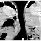
Unusual
association of parvo-virus with Morgagni hernia, mistaken for patch of consolidation. Coronal (A) and Sagittal (B) reformatted images showed herniation of LT lobe of liver along with vasculature into the RT sterno-costal space with shifting of heart towards LT side.
Morgagni-Hernie mit Hernierung des linken Leberlappens
Siehe auch:
und weiter:


 Assoziationen und Differentialdiagnosen zu Morgagni-Hernie mit Hernierung des linken Leberlappens: