multifocal fatty liver metamorphosis


Multifocal hepatic steatosis (also known as multifocal nodular hepatic steatosis) is the uncommon finding of multiple foci of focal fat in the liver mimicking - and at times being confused with - hepatic metastases.
Epidemiology
Risk factors
Conditions that increase one's risk of developing multifocal hepatic steatosis are identical to other forms of steatosis:
- diabetes mellitus
- obesity
- chronic alcohol excess
- exogenous steroids
- drugs (amiodarone, methotrexate, chemotherapy)
- IV hyperalimentation
Clinical presentation
Multifocal hepatic steatosis is usually an incidental imaging finding.
Pathology
Radiographic features
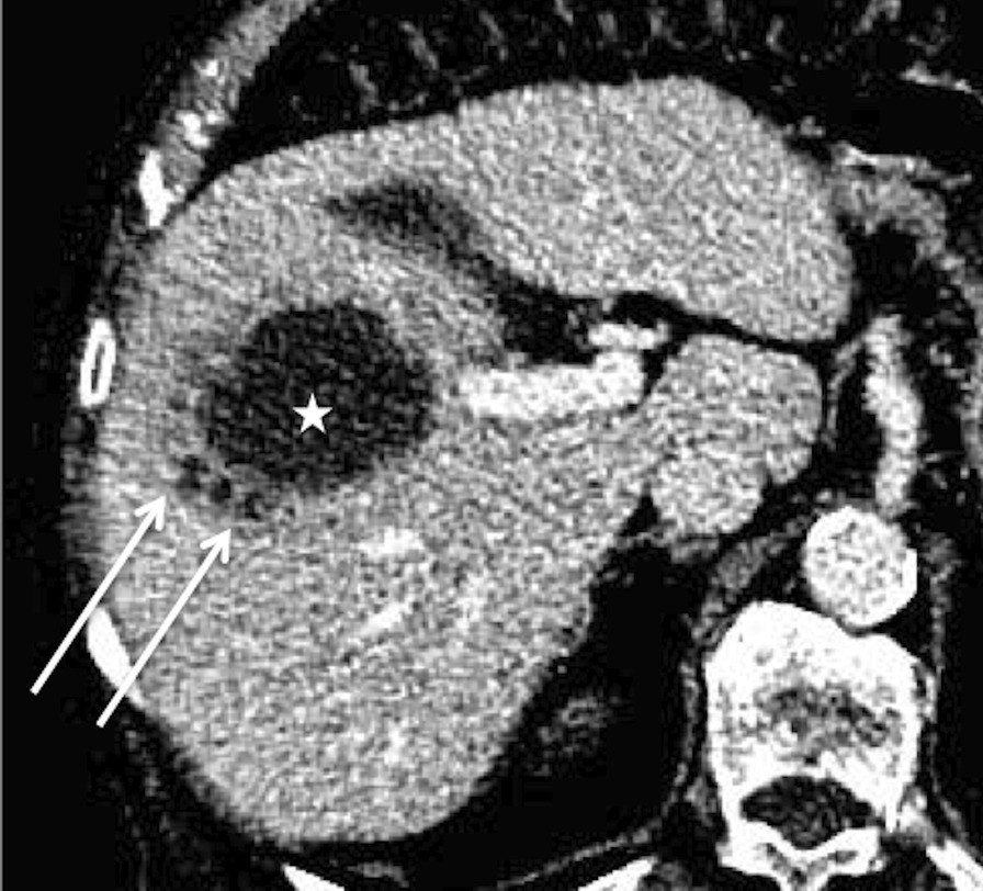
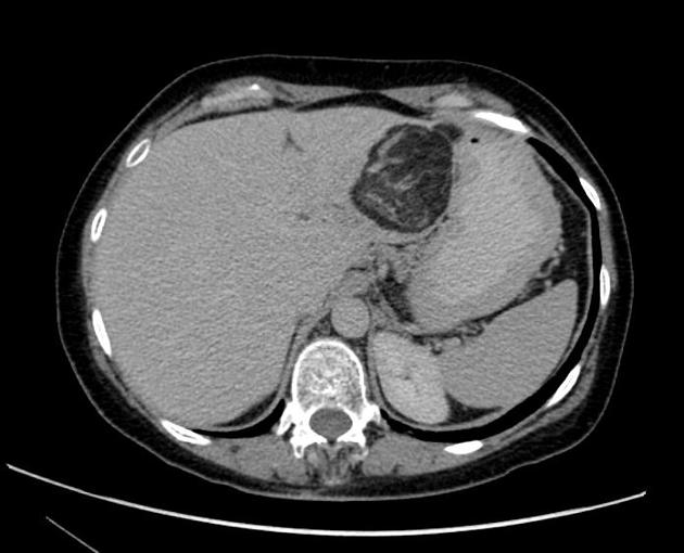
The steatotic lesions vary from several millimeters to centimeters in size . They lack mass effect (i.e. they do not displace hepatic vessels or other structures) and display no internal vascularity .
Ultrasound
Focal steatosis on ultrasound usually forms a well-circumscribed area of echogenicity without mass effect. Acoustic shadowing may be present. Using color Doppler usually shows a complete absence, or only slight flow, within the affected liver .
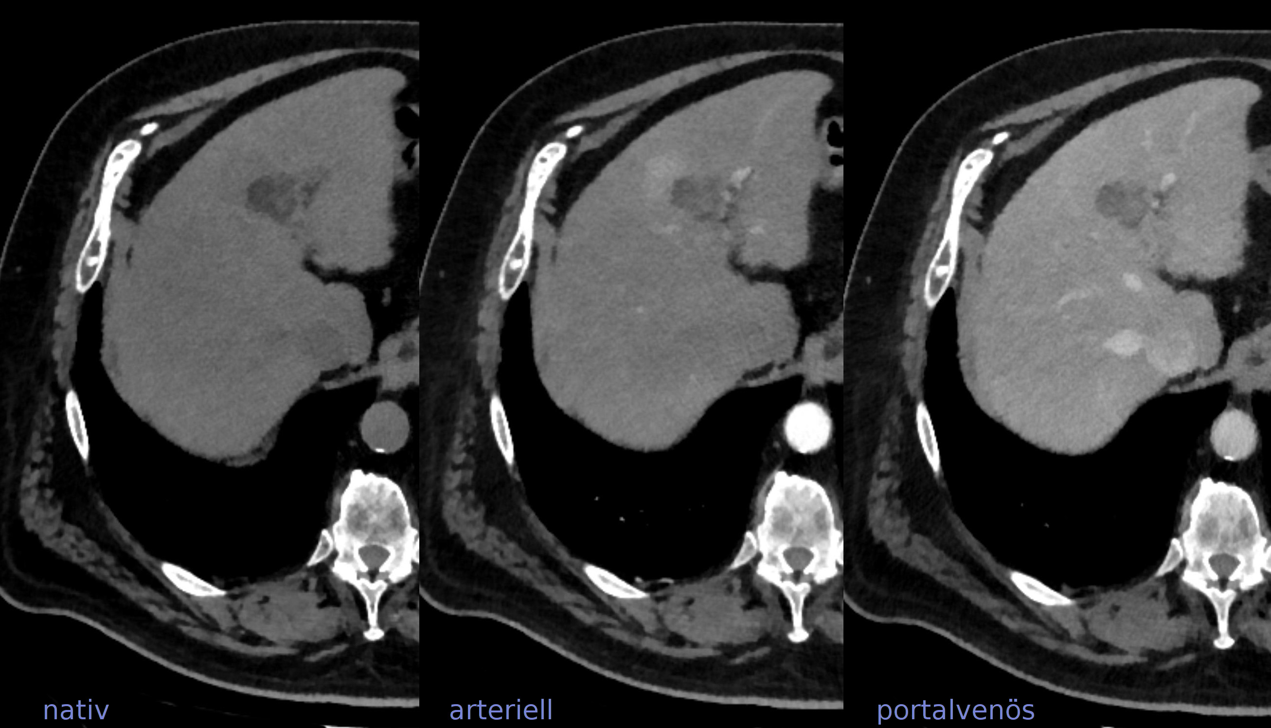
CT
Focal steatosis on CT usually forms a low density lesion without mass effect or visible enhancement.
MRI
The most discriminating finding on MRI is the loss of intralesional signal on the out-of-phase sequence.
Signal characteristics
- T1: hyperintense signal
- T2: mildly increased signal
- T1 C+ (Gd-EOB-DTPA): on the hepatobiliary phase (20 minutes), uniform enhancement of the liver across both affected and unaffected regions
- IP/OP
- in-phase: hyperintense signal within the steatotic foci
- out-of-phase: loss of signal
- DWI/ADC: no abnormal restriction is usually seen
Siehe auch:
- hypodense Leberläsionen
- Steatosis hepatis
- fetthaltige Leberläsionen
- Leberabszess
- Lebermetastasen
- Angiomyolipom der Leber
- fokale Leberverfettung
- fokale Minderverfettung der Leber
- multifokale noduläre Steatosis hepatis
und weiter:


 Assoziationen und Differentialdiagnosen zu fatty metamorphosis of the liver: