multizentrisches niedriggradiges Gliom

The
importance of fMRI and DTI in the presurgical planning of multicentric low-grade gliomas: a case report.. Coronal tractography DTI images (A,B) demonstrating the corticospinal tracts, showing thinning and displacement of the fibers on the left side.

The
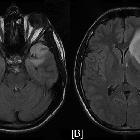
importance of fMRI and DTI in the presurgical planning of multicentric low-grade gliomas: a case report.. Axial FLAIR (Fluid Attenuated Inversion Recovery) images (A, B) reveal the high signal intensity of the two lesions located at the right temporal lobe and the left frontotemporal lobe regions respectively accompanied with ring-like oedema.

The
importance of fMRI and DTI in the presurgical planning of multicentric low-grade gliomas: a case report.. Axial FA colour-mapping images (A-D). The tumours, located at the right temporal and left frontotemporal regions, are visualized as a well-circumscribed bluish area, which is characteristic of low fractional anisotropy.

The
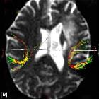
importance of fMRI and DTI in the presurgical planning of multicentric low-grade gliomas: a case report.. DTI tractography images (A-C) demonstrating the right and left arcuate fasiculli. The left one is not invaded but slightly displaced, while the right one is completely unaffected by the tumour.

The
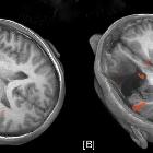
importance of fMRI and DTI in the presurgical planning of multicentric low-grade gliomas: a case report.. Presurgical fMRI images(A) showing functional language areas on the left temporoparietal lobe posterior to the tumour. These areas remain almost untouched after the surgical excision of the tumour (B).

The
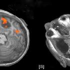
importance of fMRI and DTI in the presurgical planning of multicentric low-grade gliomas: a case report.. Presurgical fMRI image (A) showing functional language areas (Wernicke) on both temporal lobes in contact with the tumours. Postsurgically (B) the right functional area is shifted posterior to the porencephally.

 Assoziationen und Differentialdiagnosen zu multizentrisches niedriggradiges Gliom: