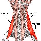
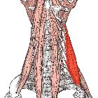
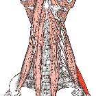
Musculi scaleni


A
cross-section diagram of the human neck at the level of C6 showing the fascia compartments, muscles, organs, bone, and major arteries, veins, and nerves.

406 Anterior
Wall of the Thorax Observe: 1. The muscles to be removed before dissection is begun: External Oblique, Rectus Abdominis, Pectoralis Major, Pectoralis Minor, Subclavius, sternal head of Sternomastoid, and Scalenus Posterior at the front; Scrratus Anterior and Latissimus Dorsi at the sides (figs. 23 & 24); Serrati Posteriores, Longissimus, and Ilio-costalis at the back (fig. 409). 2. Pulling the arm backwards and downwards forces the clavicle, padded with Subclavius, to compress the subclavian vessels against the 1st rib and thus arrest the blood flow. 3. The 11th and 12th ribs are too short to be seen from the front. 4. The 7th costal cartilages are usually the last to reach the sternum, although not uncom- monly, as here, the 8th also do so. 5. The H-shaped cut made through the perichondrium of the 3rd and 4th cartilages prior to shelling out segments of cartilage. 6. The internal thoracic vessels, crossed anteriorly by the intercostal nerves and associated with parasternal lymph nodes.


 Assoziationen und Differentialdiagnosen zu Musculi scaleni:
