ossifizierendes Fibrom des Schädels

Radiological
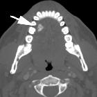
review of skull lesions. Ossifying fibroma. Water view skull radiograph (a), axial (b) and sagittal (c) head CT images show a lytic lesion in the frontal bone (arrowhead) with barely perceptible anterior and posterior cortical margins (dashed arrows)


 Assoziationen und Differentialdiagnosen zu ossifizierendes Fibrom des Schädels: