Osteomyelitis der Ferse

Imaging of
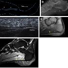
plantar fascia disorders: findings on plain radiography, ultrasound and magnetic resonance imaging. Heel osteomyelitis. Lateral plain radiograph shows marked morphological alteration of the heel with irregular lytic areas and concomitant PF thickening (double-head arrow) due to spreading of the infection (a). MRI confirms morphological alterations of the heel and PF (double-head arrow) on both T1-weighted (b) and fluid-sensitive (c) images
Osteomyelitis der Ferse
Siehe auch:


 Assoziationen und Differentialdiagnosen zu Osteomyelitis der Ferse: