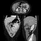
peritoneales Lymphom

Peritoneal
deposits: PET/CT the keen observer. A–D Middle age male, diagnosed with small bowel lymphoma, received CTH and was referred for re-assessment with PET CT. Conventional CT images A, B show large omental cakes occupying almost the whole abdominal cavity (white arrows). Fused images C, D revealed these lesions to be FDG avid (blue arrows). Metabolically inert ascites are also noted (brown arrow)

Abdominal
Lymphoma. At stomach level T2, diffusion weighted and fusion images show marked restriction in gastric wall lesion, and also two small hepatic lesions with the same signal intensity, suggesting metastatic disease.

Abdominal
Lymphoma. At stomach level T2, diffusion weighted and fusion images show marked restriction in gastric wall lesion, and also two small hepatic lesions with the same signal intensity, suggesting metastatic disease.
peritoneales Lymphom
Siehe auch:
und weiter:

 Assoziationen und Differentialdiagnosen zu peritoneales Lymphom:


