A radiologically inserted gastrostomy (RIG) is a procedure where a tube is inserted percutaneously in the stomach, principally to provide nutritional support for patients with swallowing disorders .
Indications
- inadequate oral intake due to dysphagia (neurologic disorder, oesophagal obstruction, head & neck masses)
- oesophagal leak
- decompression from gastric content
- gastroparesis
Contraindications
Absolute contraindications
Some generally accepted absolute contraindications are :
- severe coagulopathy
- dangerous percutaneous access to stomach (e.g. interposed colon)
Relative contraindications
- massive ascites
- gastric varices
- infection or neoplasia on the percutaneous tract
- prior gastric surgery
- severe gastro-esophageal reflux
Procedure details
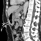
Prior to the procedure, review prior imaging studies to chose the best percutaneous approach. The patient should be NPO for the past 8 hours .
place a nasogastric tube
administer conscious sedation
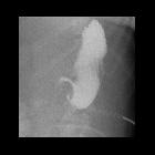
insufflate air in the nasogastric tube to distend the stomach. Enough air should be used to distend the stomach and oppose the gastric wall to the anterior abdominal wall (about 250-500cc).
choose the puncture site in the body of the stomach, equidistant from the lesser and greater curvatures
infiltrate local anesthesia with about 10cc of 1% lidocaine on the percutaneous tract
make a small skin incision with a No. 11 scalpel
use a gastropexy device (eg. a 17G needle preloaded with a Cope suture anchor) to puncture the stomach. One should feel and see the anterior stomach wall tenting upon pressure, then a give a sharp jab to enter the gastric lumen
confirm correct location by aspiration of air in a syringe
advance a stylet in the gastropexy device, to discharge the anchor in the stomach
the stylet and the gastropexy needle are then removed and the stomach is approximated to the anterior abdominal wall by a gentle traction
use your needle to make another gastric puncture, which will serve to put the gastrostomy tube. For simple gastrostomy, one should aim vertically down or slightly towards the fundus. However, if conversion to a gastrojejunostomy is anticipated, the needle should be directed towards the pylorus to have less resistance
confirm intraluminal localization with a small amount of contrast, which will outline gastric folds
insert a 0.038-inch J-tipped stiff guidewire and loop it in the stomach, and withdraw the needle
introduce serial fascial dilatators over the guidewire to dilate the muscular layers of the stomach, while taking care to keep tension on the anchor and to keep the guidewire inside the stomach lumen
insert the selected gastrostomy tube over the wire, using a peel-away sheath, which is then peeled
inflate the gastrostomy balloon with the required amount of sterile water (eg. 5-10cc)
confirm the proper position of the tube, then apply a small tension on the gastric wall by pulling the tube, while applying the retention disc on the skin, which will fix the anterior gastric wall to the anterior abdominal wall
fix the retention disc on the skin with a non-resorbable suture
Some interventional radiologists perform RIG without gastropexy, using a single puncture.
Complications
The procedure complications include :
See also
Siehe auch:


 Assoziationen und Differentialdiagnosen zu perkutane radiologisch kontrolliert platzierte Gastrostomie: