Plantar fascial tear

Imaging of
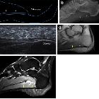
plantar fascia disorders: findings on plain radiography, ultrasound and magnetic resonance imaging. Plantar fascia rupture. On ultrasound, a tear in the PF (arrow) is shown; the PF is hypoechoic and thickened as a result of previous plantar fasciitis treated with local injections (a). MRI confirms PF rupture (arrow) and highlights marked oedema of soft tissues (b)
Plantar fascial tears refer to disruption of plantar fascial fibers which can occur in associated with longstanding plantar fasciitis or those treated with steroid injections. The tears can be complete (i.e. rupture) or incomplete.
Radiographic features
MRI
MR imaging features include:
- T1
- absence of T1-weighted low signal intensity at the site of complete rupture or partial loss of T1-weighted low signal intensity.
- abnormal thickening of the plantar aponeurosis at the site of disruption (either complete or partial)
- T1 C+ (Gd): there can be perifascial contrast enhancement as was depicted in the five cases of acute rupture
Siehe auch:

 Assoziationen und Differentialdiagnosen zu Plantar fascial tears:
