Plantarfasziitis und Fersensporn


Plantarfasziitis und Fersensporn
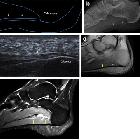
Plantarfasziitis Radiopaedia • CC-by-nc-sa 3.0 • de
Plantar fasciitis refers to inflammation of the plantar fascia of the foot. It is considered the most common cause of heel pain.
Clinical presentation
Pain on the undersurface of the heel on weight-bearing is the principal complaint. It can be worse when weight is borne after a period of rest (e.g. in the morning) and eases with walking. Passive dorsiflexion of the toes may exacerbate discomfort.
Pathology
It is generally a low-grade inflammatory process involving the plantar aponeurosis with or without the involvement of the perifascial structures.
It can arise from several factors:
- mechanical: stress of repetitive trauma (more common)
- degenerative
- systemic: as an enthesopathy in association with seronegative spondyloarthropathies
Radiographic features
Plain radiograph
Plain film features are non-specific but may show an associated plantar calcaneal spur although this is also seen in asymptomatic individuals. The thickness of the fascia can be increased to more than 4.5 mm.
Ultrasound
Often the initial imaging modality of choice. Ultrasound typically shows increased thickness of the fascia (>4.5 mm) and a hypoechoic fascia .
MRI
Signal characteristics of affected tissues include:
- T1/PD: intermediate signal
- T2: high signal
- STIR: very sensitive in the detection of both fascial and perifascial edema, which appear as poorly marginated areas of high signal intensity
Other MRI features include:
- plantar fascial thickening: often fusiform and typically involves the proximal portion and extends to the calcaneal insertion
- increased T2/STIR signal intensity of the proximal plantar fascia
- edema of the adjacent fat pad and underlying soft tissues
- limited marrow edema within the medial calcaneal tuberosity may also be seen
Treatment and prognosis
Management options are usually conservative. Local injection of steroids +/- local anesthetic may be useful to manage symptoms. Ultrasound-guided steroid injection has been shown to be effective in short-term (four-week) pain relief with reduced thickness of the plantar fascia at three months . A posterior tibial nerve block can be performed prior for a less painful plantar fascia injection.
Specific plantar fascia stretching exercises performed daily have been shown to reduce short-term (8 weeks) and long-term (2 years) pain .
Other supportive measures include weight reduction in obese patients, rest, non-steroidal anti-inflammatory drugs (NSAIDs) and reduction of weight-bearing pressure (soft rubber heel pad, molded orthosis, or heel cup or soft-soled shoes).
See also
plantarer Fersensporn Radiopaedia • CC-by-nc-sa 3.0 • de
Plantar calcaneal spurs, or sometimes simply referred to as calcaneal spurs, are a commonly seen finding in radiology practice.
Epidemiology
Plantar calcaneal spurs tend to usually occur in older men and women and may be related to obesity, osteoarthritis and current or previous heel pain.
Pathology
Plantar calcaneal spurs are thought to be a result of enthesophytic changes involving the origin of the plantar aponeurosis . Their exact pathophysiology is not well understood but many theories have been proposed .
Very rarely, plantar calcaneal spurs can fracture .
Associations
- plantar fasciitis
- adductor digiti minimi atrophy
- reactive arthritis (often ill-defined)
- psoriatic arthritis
Radiographic features
Classically seen on plain radiographs, CT and MRI as a bony spur on the sagittal image projecting inferomedially from the calcaneus.
History and etymology
Osseous spurring of the plantar aspect of the calcaneus was first documented in 1900 by the German physician P Plettner, who coined the term Kalkaneussporn (calcaneal spur) .
Siehe auch:


 Assoziationen und Differentialdiagnosen zu Plantarfasziitis und Fersensporn: