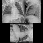
Pneumoperikard


Pneumopericardium represents gas (usually air) within the pericardium, thus surrounding the heart.
Pathology
Etiology
Underlying causes include:
- positive pressure ventilation
- thoracic surgery/pericardial fluid drainage
- penetrating trauma
- blunt trauma (rare)
- infectious pericarditis with gas-producing organisms
- fistula
- between the pericardium and an adjacent air-containing organ (i.e. stomach or esophagus)
Radiographic features
Plain radiograph and CT
On both chest radiographs and CT, appearances are characteristic, the heart being partially or completely surrounded by gas, with the pericardium sharply outlined by gas density on either side. Continuous diaphragm sign may be present.
Ultrasound
The introduction of a smooth air-soft tissue interface when intra-pericardial air is present results in horizontal reverberation artifacts obscuring the heart, appearing identical to A-lines. The appearance is similar to pneumomediastinum, but may be differentiated by the following features;
- heart remains obscured throughout the cardiac cycle
- involves the subxiphoid window
- characteristically spared in pneumomediastinum
Treatment and prognosis
Complications
Differential diagnosis
A pneumopericardium can usually be distinguished from pneumomediastinum since gas in the pericardial sac should not rise above the anatomic limits of the pericardial reflection on the proximal great vascular pedicle. Also on radiographs obtained with the patient in the decubitus position, gas in the pericardial sac will shift immediately, while gas in the mediastinum will not shift in a short interval between films.
Occasionally, it may not be possible to distinguish pneumopericardium from pneumomediastinum on plain radiographs.
Siehe auch:
und weiter:

 Assoziationen und Differentialdiagnosen zu Pneumoperikard:
