Refsum-Syndrom

Magnetic
Resonace Imaging findings in a case of infantile Refsum disease. FLAIR axial image (a) shows patchy periventricular white matter hyperintensities with sparing of the U-fibres.
Magnetic
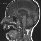
Resonace Imaging findings in a case of infantile Refsum disease. Sagittal T1-weighted image (b) reveals cortical atrophy and thinning of the body of the corpus callosum.
Magnetic
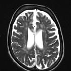
Resonace Imaging findings in a case of infantile Refsum disease. Axial and coronal (c-d) T2-weighted images confirm white matter changes in the supratentorial periventricular areas and in the cerebellum.
Magnetic
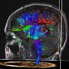
Resonace Imaging findings in a case of infantile Refsum disease. 3D sagittal presentation of fibres confirms reduction of the blue fibres in the rolandic areas and posterior corona radiata, arrangement of the fibere is relatively conserved in the anterior tracts.
Magnetic
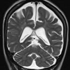
Resonace Imaging findings in a case of infantile Refsum disease. Axial and coronal (c-d) T2-weighted images confirm white matter changes in the supratentorial periventricular areas and in the cerebellum.
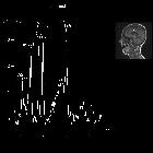
Magnetic
Resonace Imaging findings in a case of infantile Refsum disease. Spectroscopy evaluation at the level of the corona radiata reveals increased Cholin (Cho)/N-acetylaspartate (NAA) ratio and increased Myo-inositol (Mio), Lipid (Lip) and lactate (Lac) peaks.
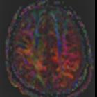
Magnetic
Resonace Imaging findings in a case of infantile Refsum disease. DTI directional map at the level of the corona radiata shows reduction of blue fibres and increased red ones in the posterior corona radiata, arrangement of the fibres is relatively conserved in the anterior tracts.
 Assoziationen und Differentialdiagnosen zu Refsum-Syndrom:


