Riesenzellependymom

Immunohistochemical
features of giant cell ependymoma of the filum terminale with unusual clinical and radiological presentation. Neuroimaging findings of the GCE from filum terminale. a-d Axial gadolinium-enhanced L3-L5-weighted CT image demonstrated an intradural non-encapsulated heterogeneously enhanced solid mass attached to the filum terminale

Immunohistochemical
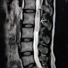
features of giant cell ependymoma of the filum terminale with unusual clinical and radiological presentation. Sagittal MRI showed a large infiltrative mass in the filum terminale. a Hyperintensity MRI T2-weighted sequence; and (b) Gadolinium-enhanced MRI scan showed a large infiltrative mass with heterogeneous high signals


 Assoziationen und Differentialdiagnosen zu Riesenzellependymom: